Chromosomal translocation
Chromosomal translocation alters genome structure, affecting health.

DataBase Center for Life Science (DBCLS) · CC BY 4.0
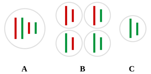
Chromosomal translocation is a genetic phenomenon involving the rearrangement of chromosome segments. It includes balanced and unbalanced forms, with three main types: reciprocal, nonreciprocal, and Robertsonian translocation. These structural changes can lead to disorders in growth, function, and development, often due to alterations in the genome.
- field
- Genetics
- known_for
- Chromosomal rearrangement causing genetic disorders and cancer
Lore & Background
Chromosomal translocations were first observed in the early 20th century. In 1916, William R. B. Robertson documented a chromosomal fusion in grasshoppers, now known as Robertsonian translocation. In 1938, Karl Sax demonstrated that X-ray irradiation could induce chromosomal translocations in plant cells. During the 1940s, Barbara McClintock's maize cytogenetics experiments revealed the breakage–fusion–bridge cycle, further illuminating mechanisms of chromosomal rearrangement. A major breakthrough came in 1960 with the discovery of the Philadelphia chromosome in chronic myelogenous leukemia, the first consistent chromosomal abnormality linked to a human cancer. In 1973, Janet Rowley identified the Philadelphia chromosome as a translocation between chromosomes 9 and 22, definitively linking a specific translocation to leukemia.
Reader's Guide
Chromosomal translocations are significant because they can cause genetic disorders, infertility, miscarriages, and cancers. Balanced reciprocal translocations are usually harmless but can lead to unbalanced gametes, resulting in conditions like Emanuel syndrome. Unbalanced translocations cause duplication or deletion of genetic material, often leading to phenotypic abnormalities. Robertsonian translocations, involving acrocentric chromosomes, can result in conditions such as translocation Down syndrome. Technological advances, including chromosome banding in the 1970s, fluorescence in situ hybridization (FISH) in the 1980s, and high-throughput DNA sequencing in the 21st century, have greatly enhanced detection and understanding. These methods allow precise mapping of breakpoints and discovery of translocations across cancers and genetic disorders. The legacy of translocation research includes improved genetic counseling and testing for families, as well as deeper insight into the role of chromosomal rearrangements in human disease.
Did You Know?
- The Philadelphia chromosome, discovered in 1960, was the first consistent chromosomal abnormality linked to a human cancer.
- Robertsonian translocation was first documented in grasshoppers by William R. B. Robertson in 1916.
- Carriers of Robertsonian translocations involving chromosome 21 have a higher risk of having a child with Down syndrome.
- Fluorescence in situ hybridization (FISH), developed in the early 1980s, uses fluorescent probes to map translocation breakpoints.
The Architecture of Chromosomal Rearrangement
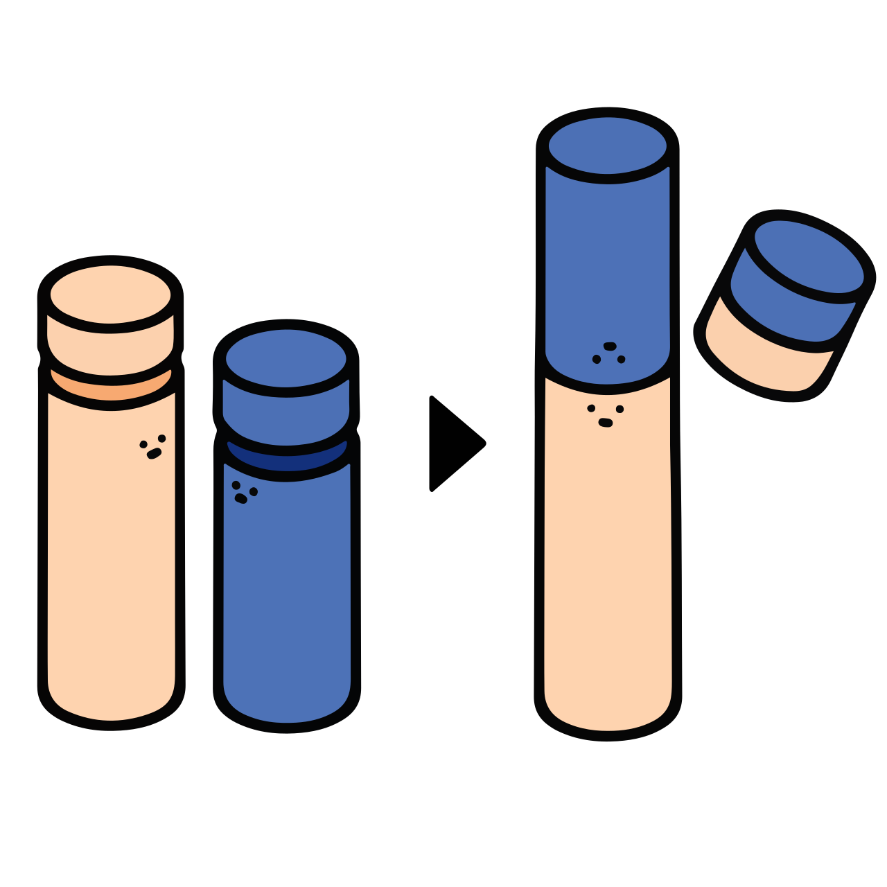
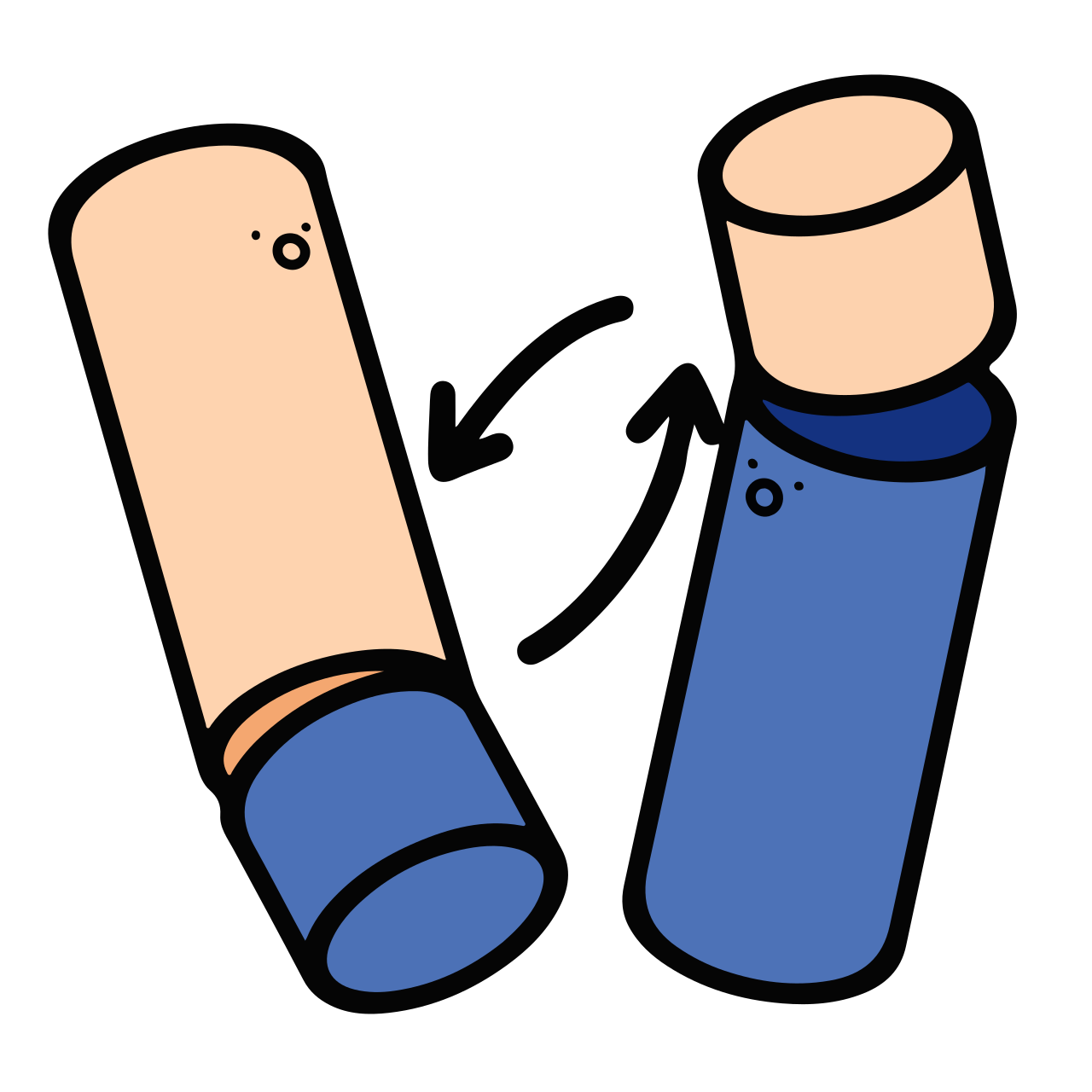
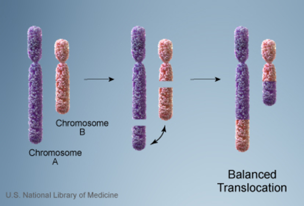
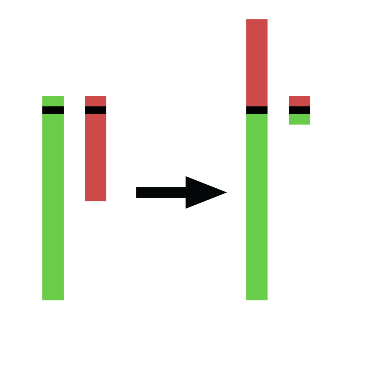
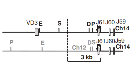
Chromosomal translocation represents a fundamental disruption in the orderly arrangement of genetic material, manifesting in three principal forms: reciprocal, nonreciprocal, and Robertsonian. In reciprocal translocation, two non-homologous chromosomes swap detached fragments, effectively trading segments between themselves. Robertsonian translocation takes a different approach, where one chromosome from each of two healthy pairs fuses together into a single blended structure. Nonreciprocal translocation involves a one-way transfer, where material moves from a donor chromosome to an acceptor without any reciprocal exchange. These rearrangements can be classified as balanced, where no genetic material is gained or lost and full functionality is theoretically preserved, or unbalanced, where the exchange produces extra or missing genes. A particularly consequential outcome occurs when a translocation bridges two genes that were previously separated, creating a gene fusion. The underlying structural changes—deletions, duplications, and inversions—ultimately shape the developmental and functional trajectory of the affected individual.
A Century of Discovery: From Grasshoppers to the Genome
The story of chromosomal translocation spans over a century of biological inquiry. In 1916, American zoologist William R. B. Robertson first documented a chromosomal fusion in grasshoppers, a finding that would later bear his name as Robertsonian translocation. Decades later, Karl Sax demonstrated in 1938 that X-ray irradiation could force fusions between different chromosomes in plant cells, establishing a causal link between external damage and rearrangement. Barbara McClintock's maize cytogenetics work in the 1940s further illuminated the breakage-fusion-bridge cycle, deepening understanding of how chromosomes rearrange. The field's most transformative moment arrived in 1960 with the identification of the Philadelphia chromosome in chronic myelogenous leukemia, the first consistent chromosomal abnormality tied to a human cancer. In 1973, Janet Rowley pinpointed this as a translocation between chromosomes 9 and 22, cementing the connection between a specific rearrangement and a specific disease. These milestones laid the groundwork for modern genomic medicine.
Balanced Carriers and the Fertility Burden
Balanced reciprocal translocations, in which two non-homologous chromosomes exchange material in an even trade, typically leave the carrier free of visible phenotypic effects. A well-known example involves the exchange of segments between chromosomes 11 and 22; individuals carrying this rearrangement appear healthy yet face significant reproductive challenges. The danger emerges during meiosis, when the carrier's chromosomes segregate unevenly, producing gametes that carry unbalanced reciprocal or nonreciprocal translocations. When such a gamete unites with a normal gamete from the other parent, the resulting embryo may harbor duplications or deletions of genetic material. The clinical consequences range from infertility and recurrent miscarriages to children born with structural abnormalities. Emanuel syndrome stands as a recognized birth defect that can arise from a carrier of the 11;22 translocation. Because of these risks, genetic counselling and targeted genetic testing are routinely offered to families in which a balanced translocation has been identified, providing prospective parents with information about the probability and nature of potential outcomes.
From Banding Patterns to Single-Nucleotide Resolution
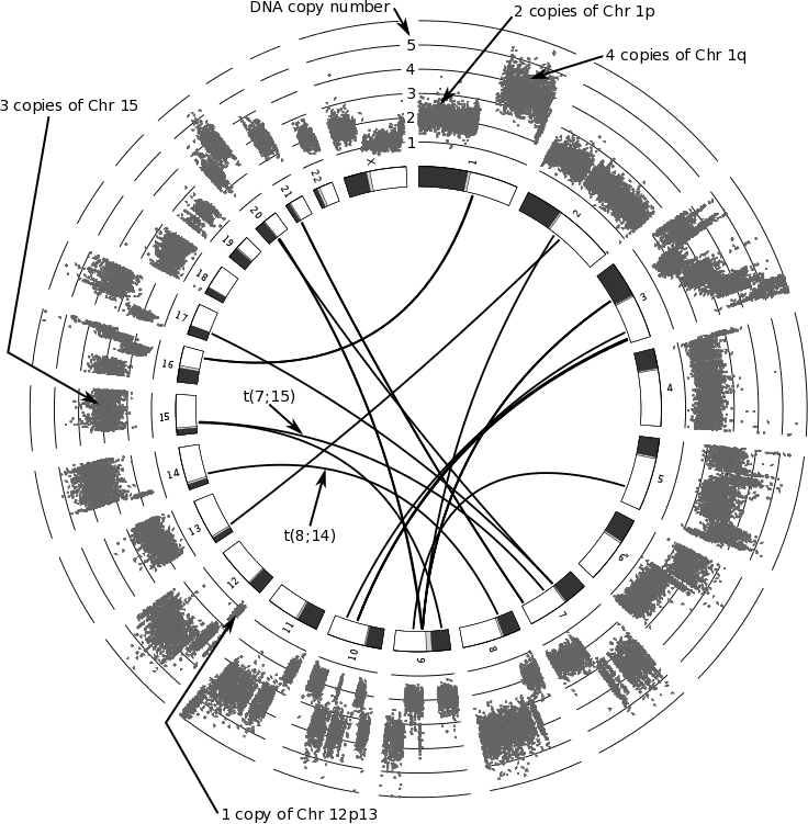
Detecting chromosomal translocations has evolved dramatically alongside advances in laboratory technology. Early identification relied on cytogenetics and karyotyping of affected cells, where researchers visually inspected chromosome structure. The 1970s brought chromosome banding techniques such as Q-banding and G-banding, which produced distinctive striped patterns on each chromosome, enabling far more precise identification of individual chromosomes and their structural abnormalities. A quantum leap came in the early 1980s with fluorescence in situ hybridization, or FISH, which allowed scientists to attach fluorescent probes to specific DNA sequences directly on chromosomes, dramatically sharpening the mapping of translocation breakpoints. In the twenty-first century, high-throughput DNA sequencing methods, including whole-genome sequencing, have pushed detection to single-nucleotide resolution. This capability has uncovered numerous translocations that were previously invisible, expanding the catalog of rearrangements associated with various cancers and genetic disorders and opening new avenues for diagnosis and targeted therapeutic strategies.
Gallery
More in Genetics 1-24
Spotted an error? Know more?
This is a living reference — every entry is fact-audited, and reader corrections feed straight into our audit queue. Suggest an edit · See this site's audit record