Chromatin
DNA–protein complex that packages and regulates genetic material.

Courtesy of NIAID Ryan Kissinger · Public domain
Chromatin is the combination of DNA and proteins that compresses and organizes chromosomal DNA. This structure appears in both bacterial and eukaryotic cells. In eukaryotes, chromatin is mainly made up of DNA attached to histone proteins, along with various other chromatin-binding factors that help organize and regulate the genome. Its job is to pack long DNA molecules into compact forms while managing access to genetic information for tasks like transcription, DNA replication, and DNA repair. When cells divide, chromatin ensures chromosomes separate correctly during anaphase; the distinct shapes of chromosomes seen at this stage come from DNA being tightly coiled into highly condensed chromatin.
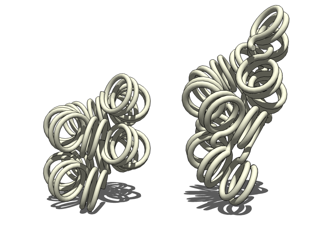
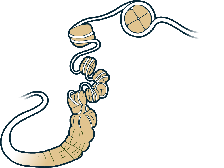
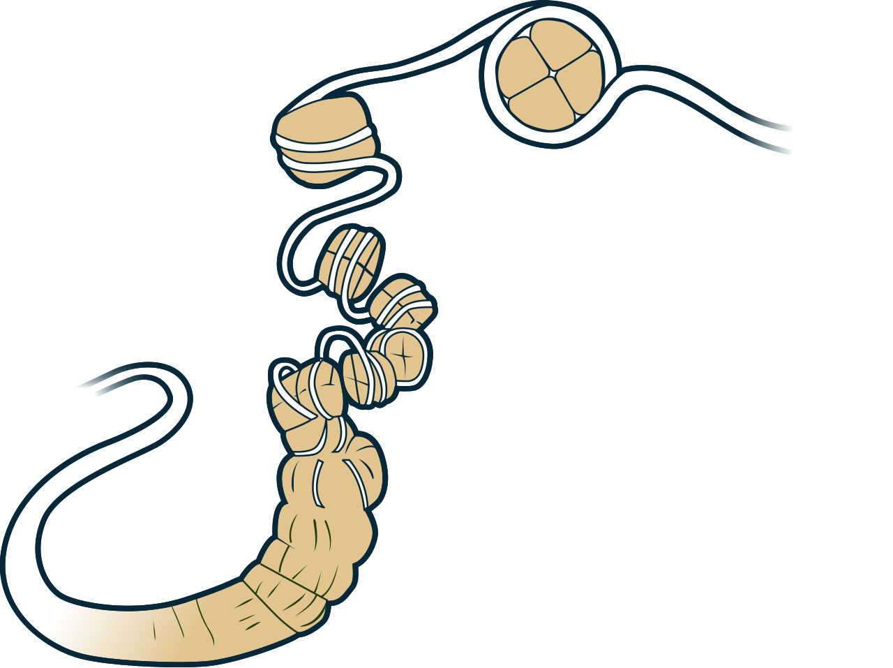
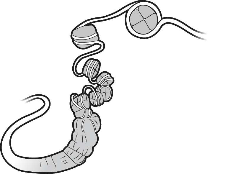
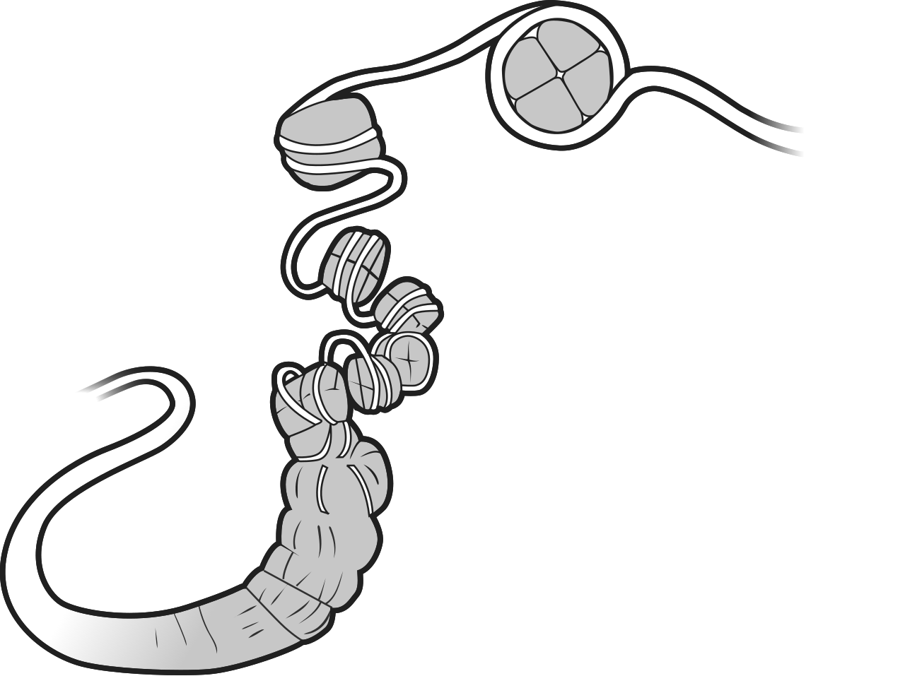
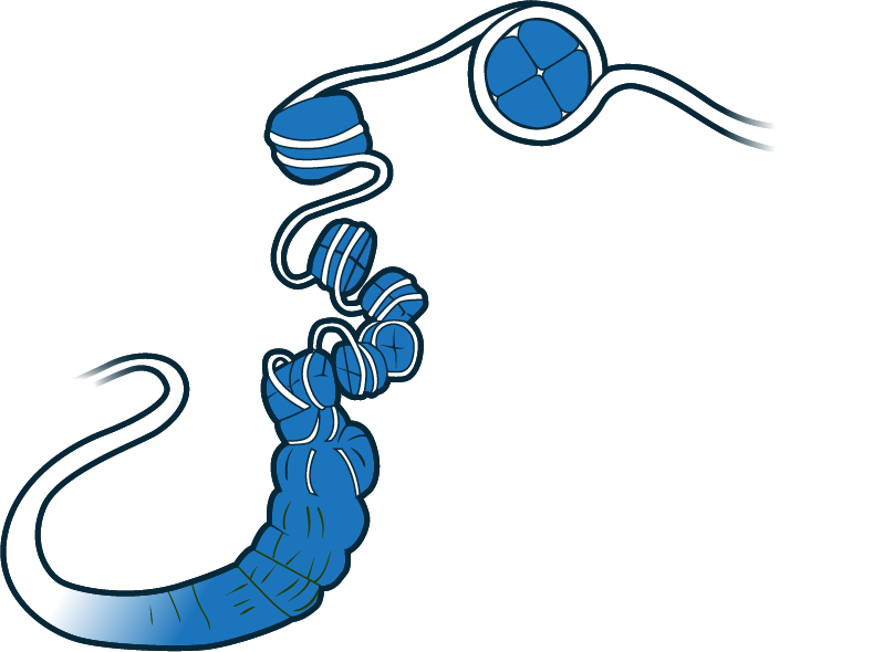
The organization of chromatin is usually described at several structural levels. At the simplest level, DNA coils around histone octamers to form nucleosomes, which are linked by stretches of linker DNA. This creates a “beads-on-a-string” fiber roughly 10–11 nm wide. These nucleosome arrays can interact with each other and with linker histones to build more complex structures. A 30-nm chromatin fiber has been seen in lab experiments, but whether it actually exists in living cells is still debated.
On a larger genomic scale, chromatin forms loops and domains that shape the genome’s three-dimensional layout. Chromosomes are split into compartments linked to active (euchromatin) or inactive (heterochromatin) states, and each chromosome occupies its own distinct area in the nucleus, called a chromosome territory.
Chromatin organization varies across organisms. For instance, sperm cells and avian red blood cells pack their chromatin much more tightly than most eukaryotic cells. On the other hand, some protozoa, like trypanosomatids, don’t condense their chromatin into visible chromosomes at all. Bacteria organize their DNA differently, forming a chromatin or nucleoid structure controlled by nucleoid-associated proteins such as H-NS and StpA. Some archaea have histone proteins and package DNA into nucleosome-like assemblies of varying sizes, sometimes called hypernucleosomes.
Chromatin structure also changes throughout the cell cycle. During interphase, chromatin is generally looser, giving RNA and DNA polymerases access to transcribe and replicate DNA. The local structure of interphase chromatin depends on the specific genes present. In mitosis and meiosis, chromatin
- field
- Molecular biology, genetics
- known_for
- Packaging DNA into nucleosomes and higher-order structures; regulating gene expression
- components
- DNA, histones (H2A, H2B, H3, H4, H1), linker DNA, nucleosome core particle
- structural_levels
- 10–11 nm beads-on-a-string fiber, debated 30-nm fiber, loops, TADs, A/B compartments, chromosome territories
Lore & Background
The primary protein components of chromatin are histones. The nucleosome, the fundamental basic unit of chromatin, consists of DNA wrapped around a histone octamer containing two copies each of the core histones H2A, H2B, H3, and H4. Approximately 147 base pairs of DNA wrap around this octamer to form the nucleosome core particle. Neighboring nucleosomes are connected by stretches of linker DNA, which vary in length among organisms and cell types but typically range from about 20 to 60 base pairs. Arrays of nucleosomes connected by linker DNA form an extended fiber often described as a 'beads-on-a-string' structure approximately 10–11 nm in diameter under low-salt or experimentally reconstituted conditions. In addition to core histones, a linker histone H1 binds near the entry and exit sites of DNA on the nucleosome and contributes to higher-order chromatin organization.
Reader's Guide
Chromatin organization is often described at several structural levels. At the most basic level, DNA wrapped around histone octamers forms nucleosomes connected by stretches of linker DNA. Under certain experimental conditions, nucleosome arrays can fold into more compact structures with diameters of approximately 30 nm, though the existence of a uniform 30-nm chromatin fiber in living cells remains debated. At larger genomic scales, chromatin is organized into loops and domains that contribute to the three-dimensional architecture of the genome. Chromosomes are further partitioned into compartments associated with active (euchromatin) or inactive (heterochromatin) chromatin states, and individual chromosomes occupy distinct spatial regions within the nucleus known as chromosome territories. Chromatin structure is highly dynamic and changes throughout the cell cycle and in response to cellular signals. Histone modifications, including acetylation and methylation, alter local chromatin structure and therefore gene expression.
Did You Know?
- Chromatin is found in both bacterial and eukaryotic cells.
- The nucleosome core particle, together with histone H1, is known as a chromatosome.
- Some protozoa such as trypanosomatid do not condense their chromatin into visible chromosomes at all.
- Histone acetylation is generally correlated with increased chromatin accessibility and active transcription.
The Nucleosome: Chromatin's Fundamental Building Block
The smallest repeating unit of eukaryotic chromatin is the nucleosome, a structure in which roughly 147 base pairs of DNA spiral around a protein core called a histone octamer. This octamer is assembled from two copies each of four core histones—H2A, H2B, H3, and H4—creating a compact platform that tethers the long DNA molecule in place. Neighboring nucleosomes are linked by stretches of linker DNA, typically spanning 20 to 60 base pairs depending on the organism and cell type. When visualized under low-salt or reconstituted laboratory conditions, these repeating units produce the classic beads-on-a-string fiber, roughly 10 to 11 nanometers in diameter. A fifth histone, H1, docks near the entry and exit points of DNA on each nucleosome, and the combination of the core particle plus H1 is termed a chromatosome. Although nucleosomes bind DNA in a largely non-specific manner suited to general packaging, pronounced sequence preferences govern their positioning. DNA regions rich in adenine and thymine are more easily compressed into the inner minor grooves, so nucleosomes tend to lock into place at intervals matching the roughly ten-base-pair helical repeat of the double helix.
Three-Dimensional Architecture and the Loop Economy
Beyond the linear string of nucleosomes, chromatin folds into a rich three-dimensional landscape that governs how genes are read. At the genomic scale, DNA forms dynamic loops that bring distant regulatory elements into proximity with their target genes, sharpening the efficiency of transcriptional interactions. These loops are not static; they continuously form and dissolve. Two key molecular players orchestrate this process. Cohesin complexes, which resemble ring-shaped protein assemblies, generate loops by threading the DNA fiber through their central channel in a process called extrusion. CTCF, a sequence-specific transcription factor, acts as a boundary element: when two CTCF molecules are oriented in opposite directions along the DNA, they halt the advancing cohesin ring and define the edges of a loop. Additional regulators such as Jpx influence where CTCF can bind along the fiber. On a broader scale, chromosomes are partitioned into active euchromatin compartments and repressed heterochromatin compartments, and each chromosome occupies its own spatial territory within the nucleus. Chemical modifications on histone tails—methylation and acetylation among them—further tune local compaction and thus gene expression.
The 30-nm Fiber: A Model Under Scrutiny
For decades, a central tenet of chromatin biology held that the beads-on-a-string fiber folds into a regular, uniform structure roughly 30 nanometers in diameter. Early structural models envisioned this fiber as either a one-start solenoid, in which nucleosomes wind in a single helical path, or a two-start zigzag, in which nucleosomes alternate sides of a central axis. These elegant pictures dominated textbooks and research for years. However, the reality inside living cells has proven far messier. The existence of a consistent 30-nm fiber in vivo remains genuinely contested. High-resolution techniques, particularly cryo-electron microscopy, have revealed that chromatin in many cell types assembles into irregular, heterogeneous, and highly dynamic nucleosome arrangements rather than the neat periodic fibers once assumed. The 30-nm structure can still be observed under specific in vitro conditions, but its prevalence and biological relevance in the crowded, active environment of a living nucleus are no longer taken for granted. This ongoing debate underscores how much of chromatin's higher-order organization is context-dependent, shaped by the particular cell type, its metabolic state, and the genomic region under examination.
Chromatin Across Life and Through the Cell Cycle
Chromatin is not a one-size-fits-all structure; its organization shifts dramatically across species and across the phases of a single cell's life. In bacteria, DNA is packaged into a nucleoid organized by proteins such as H-NS and StpA rather than histones. Some archaeal species, however, encode histone-like proteins and assemble nucleosome-like structures of variable size, sometimes called hypernucleosomes. Among eukaryotes, spermatozoa and avian red blood cells pack their chromatin far more tightly than typical somatic cells, while certain protozoa in the trypanosomatid group never condense their DNA into visibly discrete chromosomes at all. Within a single eukaryotic cell, chromatin breathes with the cell cycle. During interphase, it remains relatively open so that RNA and DNA polymerases can access their templates, and the local degree of compaction varies gene by gene. When the cell enters mitosis or meiosis, chromatin compacts dramatically, producing the characteristic rod-shaped chromosomes that are segregated to daughter cells during anaphase. This cyclical tightening and loosening is essential for both faithful inheritance and accessible gene expression.
Gallery
More in Genetics 1-24
Spotted an error? Know more?
This is a living reference — every entry is fact-audited, and reader corrections feed straight into our audit queue. Suggest an edit · See this site's audit record