3-M syndrome
A rare genetic disorder with severe growth retardation and craniofacial abnormalities.
3-M syndrome, also known as 3M3, is a rare hereditary disorder marked by severe growth retardation, distinctive facial features, and skeletal abnormalities. It was first identified by researchers Miller, McKusick, and Malvaux, whose initials give the condition its name. The disorder is caused by mutations in one of three genes—CUL7, OBSL1, or CCDC8—and follows an autosomal recessive inheritance pattern.
Quick Facts
- First described
- 1972
- Inheritance
- Autosomal recessive
- Known cases worldwide
- Less than 100
- Genes involved
- CUL7
- OBSL1
- CCDC8
Facts from the source article.
Did You Know?
- Carriers who inherit only one mutant copy do not show symptoms of the disorder.
- Recent studies propose that CUL7, OBSL1, and CCDC8 proteins form a complex that maintains microtubule and genome integrity.
Symptoms and signs
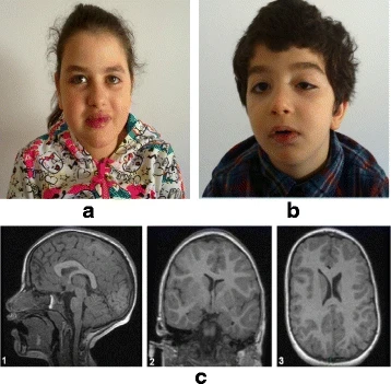
Individuals with 3-M syndrome experience severe prenatal growth retardation, resulting in low birth weight despite being carried to term. Growth delays persist after birth throughout childhood and adolescence, leading to short stature with proportional limb development. Many affected infants have distinctive craniofacial abnormalities, including premature closure of the sagittal sutures, which may cause the skull to appear abnormally long and narrow. The forehead may be prominent, the face triangular with a hypoplastic midface and pointed chin, and the cheeks flat. Other features include large ears, a prominent mouth with widely spread lips, underdeveloped upper jaw bones, and crowded teeth. Skeletal anomalies develop after birth and include delayed bone maturation, slender long bones, tall vertebral bodies, joint hypermobility, increased risk of hip dislocation, and abnormal spinal curvature such as kyphoscoliosis or hyperlordosis. Additional findings may include a short broad neck and thorax, square shoulders, flared shoulder blades, curved fifth finger, and prominent heels.
Diagnosis
Diagnosis is suggested by the presence of congenital clinical features, including low birth weight, severe growth retardation, typical facial features, and characteristic radiological findings. Fetal ultrasonography may detect growth retardation or other suggestive findings before birth. Radiographic findings, which become apparent after the first two years of life, reveal slender long bones, tall vertebral bodies that shorten over time, small pelvic bones, a broad thorax with slender ribs, and delayed bone age. Molecular genetic testing can confirm the diagnosis and identify the specific gene involved. Testing is typically ordered by likelihood of mutation: CUL7 (77.5%), OBSL1 (16%), and CCDC8 (unknown frequency). Methods include deletion/duplication analysis, targeted variant analysis, or sequence analysis of the entire coding region.
Treatment
Treatment is tailored to each individual's symptoms and may involve a team of specialists including pediatricians, orthopedists, and dentists. Management options for short stature include surgical bone lengthening or growth hormone therapy. Orthopedic techniques or surgery address skeletal abnormalities, and plastic surgery may correct certain craniofacial anomalies. Dental issues such as crowding may be treated with braces or oral surgery. Children should be monitored for growth and pubertal progress every 6–12 months until final height is achieved. Newborns should receive a hip ultrasound to screen for developmental dysplasia. Recombinant human growth hormone (r-hGH) may be tried; response is generally modest, but if a one-year trial shows reasonable response, long-term continuation is recommended. Adaptive aids and physical therapy are also options. Genetic counseling is beneficial for affected individuals and families.
More in Syndromes with Craniofacial Abnormalities
Spotted an error? Know more?
Reader corrections go straight into our review queue. Suggest an edit · How this site is sourced