Chromatophores
A living pixel in the skin, flickered by nerves to paint the ocean in a thousand shades.

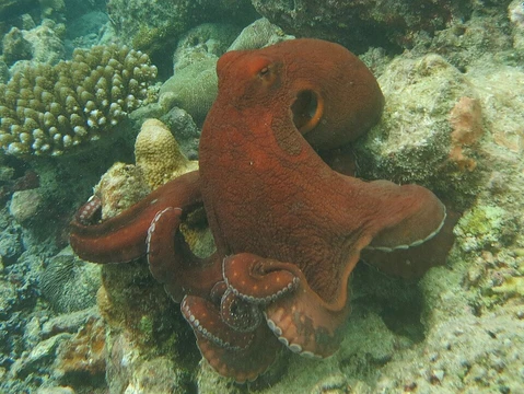
Chromatophores are pigment-bearing cells embedded in the dermis of many marine and terrestrial animals, most extensively studied in cephalopods (octopus, squid, cuttlefish) and bony fish. In cephalopods each chromatophore is a discrete, individually innervated unit: a central pigment sac surrounded by a ring of contractile muscles and elastic fibres, allowing the animal to expand or collapse the visible pigment area in milliseconds. In fish the term covers a broader family of colour cells—melanophores, xanthophores, erythrophores, iridophores, and leucophores—whose aggregation or dispersal shifts the animal's overall hue.
Their primary roles are camouflage (matching substrate or water column), intraspecific communication (mating signals, threat displays), and photoprotection. Because they sit in the outermost skin layer and are directly wired to the nervous system in cephalopods or to endocrine pathways in many fish, chromatophores represent one of the fastest and most versatile colour-change mechanisms known in the animal kingdom.
- Structure type
- Pigment-containing dermal cell
- Primary marine groups
- Cephalopods, bony fish (Teleostei), some crustaceans
- Location in body
- Dermis (outer skin layer)
- Main functions
- Camouflage, communication, photoprotection
- Control mechanism (cephalopods)
- Direct neural (motor neurons per chromatophore)
- Control mechanism (many fish)
- Hormonal (e.g., MSH, catecholamines)
- Subtypes (fish)
- Melanophores, xanthophores, erythrophores, iridophores, leucophores
Lore & Background
In the dermis of a common octopus, each chromatophore is a self-contained optical unit roughly 0.5–1 mm in diameter. At its centre sits a sac packed with eumelanin granules; radiating outward are dozens of contractile muscle fibres that pull the sac flat, while elastic fibres snap it back when the muscles relax. A single motor neuron can drive one chromatophore independently, so an octopus can produce a mosaic of expanded and contracted units across its mantle in a fraction of a second—far faster than any pigment-transport mechanism seen in vertebrate fish.
In teleost fish the picture is different. Chromatophores there are not individually innervated; instead, circulating hormones such as melanophore-stimulating hormone (MSH) or catecholamines trigger aggregation (clumping) or dispersal of pigment granules within the cell. A wrasse or damselfish shifting from a pale daytime phase to a dark nocturnal phase is essentially a whole-body redistribution of those granules, a process that takes minutes rather than milliseconds.
Cephalopod chromatophores also work in concert with iridophores—reflective guanine-crystal cells—and with papillae, which alter skin texture. The triad gives cuttlefish and octopus a three-dimensional camouflage system: colour, structural sheen, and surface roughness all change simultaneously, a level of dynamic disguise unmatched in the invertebrate world.
Reader's Guide
Pigment sac (central reservoir): A small, roughly spherical vesicle filled with eumelanin or other pigment granules. In a cephalopod it is about 0.1–0.3 mm across, a tiny fraction of the animal's body, yet it is the visible 'pixel' of the skin. Remarkable because it is individually addressable by a single motor neuron—no other invertebrate cell is controlled with that granularity.
Radial contractile muscles: A fan of dozens of muscle fibres radiating from the pigment sac to the skin surface. When they contract they flatten the sac, spreading pigment across a wider area. Their speed of contraction (tens of milliseconds) is what makes cephalopod colour change near-instantaneous.
Elastic fibres: Spring-like filaments surrounding the radial muscles. When the muscles release, elastic recoil snaps the sac back to its resting dome shape. This passive reset means the animal only expends energy to change colour, not to hold it.
Iridophores (associated structure): Guanine-crystal reflectors lying beneath the chromatophore layer. They add structural colour—silvery, blue, or green sheen—that shifts with viewing angle, giving cephalopods a dynamic, iridescent component absent in most other invertebrate skin systems.
Papillae (associated structure): Eversible dermal bumps that alter surface texture. Together with chromatophores and iridophores they let a cuttlefish mimic the rough grain of coral or sand, a three-dimensional camouflage no flat-bodied invertebrate can replicate.
Did You Know?
- In cephalopods each chromatophore is driven by its own dedicated motor neuron, so one octopus can independently expand or contract thousands of pigment units in a single frame of motion.
- The word 'chromatophore' derives from the Greek chroma (colour) and phorein (to bear or carry), literally a 'colour-bearer.'
- In many bony fish, chromatophore state is set by circulating hormones such as melanophore-stimulating hormone (MSH), meaning a colour shift can take minutes rather than the milliseconds seen in a squid.
- Cephalopod chromatophores work in a triad with iridophores (reflective guanine crystals) and papillae (textural bumps), giving a three-dimensional camouflage system unmatched among invertebrates.
More in Anatomy & Physiology
Elsewhere in Big blue octopus
Spotted an error? Know more?
Reader corrections go straight into our review queue. Suggest an edit · How this site is sourced