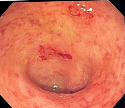
Esophagitis
Inflammation of the esophagus, often from acid reflux.

CoRus13 · CC BY-SA 4.0
Esophagitis, also spelled oesophagitis, is a disease characterized by inflammation of the esophagus. The esophagus is a muscular tube connecting the pharynx to the stomach, composed of a mucosal lining and smooth muscle fibers. It can be asymptomatic or cause symptoms such as heartburn, dysphagia, and abdominal pain, with gastroesophageal reflux disease (GERD) being the most common cause.
Quick Facts
- Complications
- Cancer
Facts from the source article.
Lore & Background
Esophagitis involves inflammation of the esophageal lining, which can result from acid reflux, infections, medications, or allergies. The lower esophageal sphincter normally prevents stomach acid from entering the esophagus; when it fails, GERD can develop. Other causes include chemical injury, physical injury from nasogastric tubes, alcohol use disorder, Crohn's disease, stress, radiation therapy, and eosinophilic esophagitis, a chronic condition with a theorized autoimmune component.
Reader's Guide
Esophagitis is significant because untreated inflammation can lead to complications such as esophageal ulcers, scarring, Barrett's esophagus, and increased risk of esophageal cancer. Diagnosis is typically made via upper endoscopy with biopsy, barium swallow, or laboratory tests. Treatment focuses on lifestyle changes—such as weight loss, smoking cessation, and avoiding trigger foods—and medications like antacids, H-2 receptor blockers, and proton pump inhibitors. For eosinophilic esophagitis, avoiding allergens and using swallowed steroids may help. The condition is more prevalent in adults and does not discriminate by gender or ethnicity.
Did You Know?
- The most common cause of esophagitis is gastroesophageal reflux disease (GERD).
- Esophagitis can be asymptomatic or cause epigastric and substernal burning pain, especially when lying down or straining.
- Infectious esophagitis cannot be spread, but infections causing it can be transmitted.
- Eosinophilic esophagitis is associated with a high concentration of eosinophils in the esophagus and may be triggered by food or inhalant allergies.
Anatomy and the Mechanics of Injury
The esophagus functions as a roughly eight-inch muscular conduit linking the throat to the stomach, propelling swallowed material through coordinated peristaltic contractions. Its wall is organized into three concentric layers: the innermost mucosa, a middle submucosa, and an outer muscularis externa. The mucosal lining itself is built from stratified squamous epithelium supported by lamina propria and a thin muscularis mucosae. At the distal end, the lower esophageal sphincter acts as a one-way gate, normally keeping gastric acid below the diaphragm. Esophagitis arises when this barrier fails or when an external irritant breaches the mucosal surface. Whether the trigger is a lax sphincter, a pathogen, a chemical burn, or a medication lodged against the wall, the result is inflammation of one or more tissue layers. That swelling narrows the lumen, turning a routine swallow into a struggle and, in severe cases, causing a food bolus to become trapped against the constricted segment.
A Wide Spectrum of Triggers
Although gastroesophageal reflux disease remains the single most frequent culprit—stomach acid washing backward through a poorly sealed sphincter to scorch the mucosa—the list of triggers is remarkably broad. Certain medications, if taken without adequate liquid or if they lodge against the esophageal wall, can erode tissue directly; NSAIDs such as aspirin, naproxen, and ibuprofen, antibiotics including doxycycline and tetracycline, quinidine, bisphosphonates prescribed for osteoporosis, corticosteroids, potassium chloride, and even iron or vitamin C supplements all figure on that list. Chemical burns from alkaline or acidic solutions, mechanical trauma from nasogastric tubing, and the repeated acid exposure of forceful vomiting represent additional pathways. Systemic conditions play a role as well: Crohn's disease can extend its inflammatory attack into the esophageal lining, chronic alcohol use progressively wears the mucosa down, and immunodeficiency opens the door to fungal, viral, or bacterial invasion. Stress elevates acid output, radiation therapy suppresses immune defenses, and food or inhalant allergies can provoke an eosinophil-rich inflammatory response. In short, esophagitis is less a single disease than a shared endpoint reached by many different routes.
Diagnosis and the Taxonomy of Subtypes
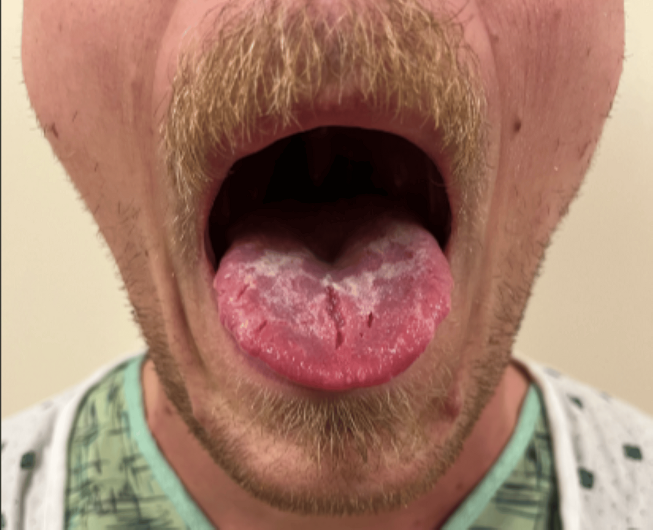
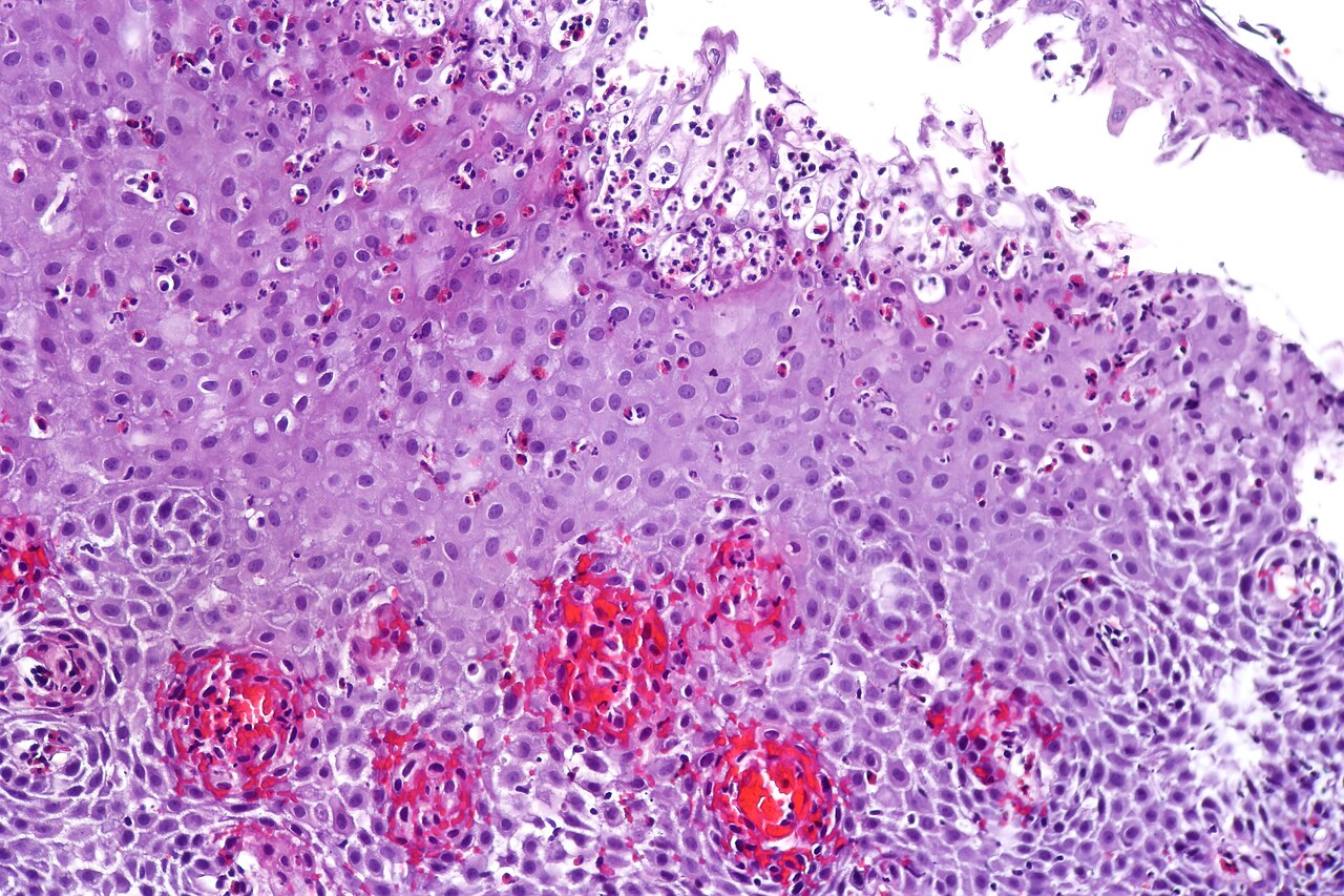
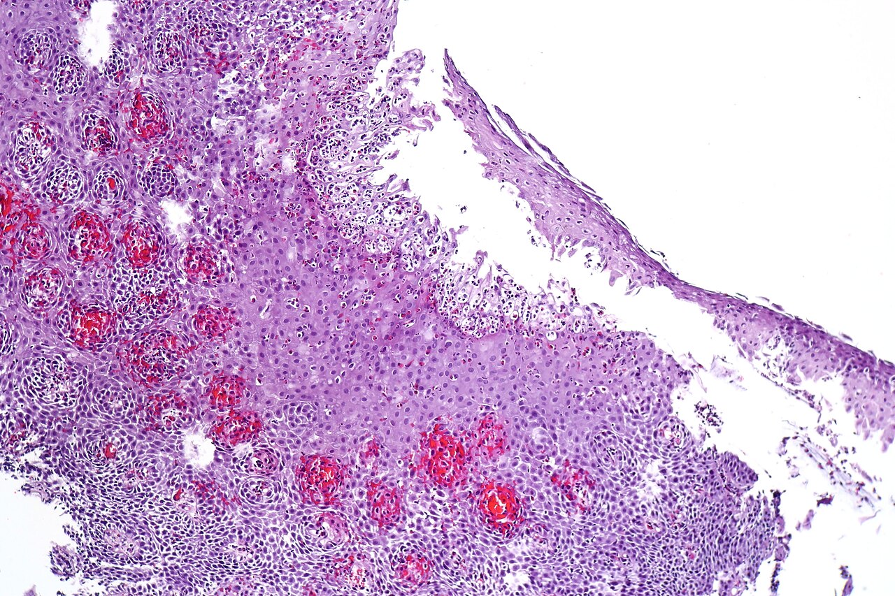
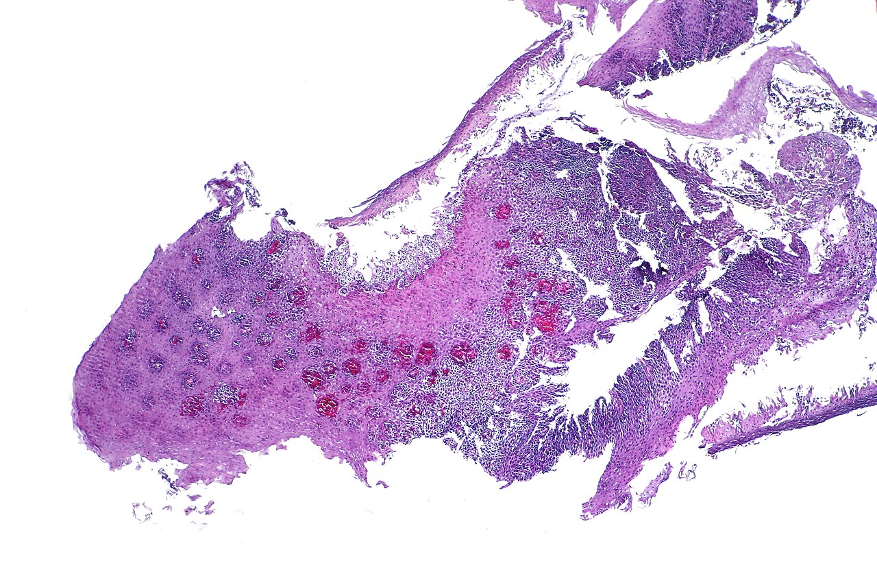
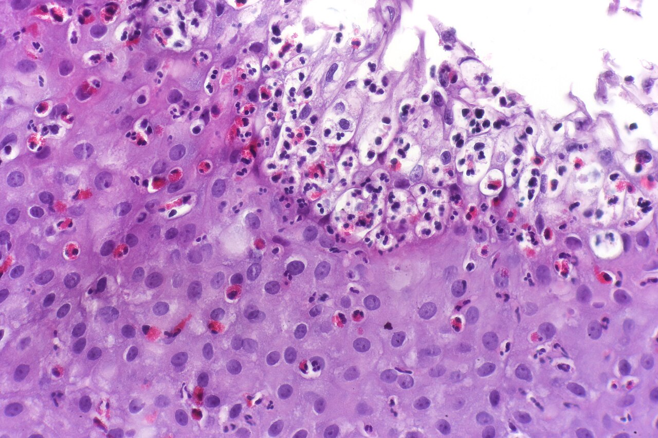
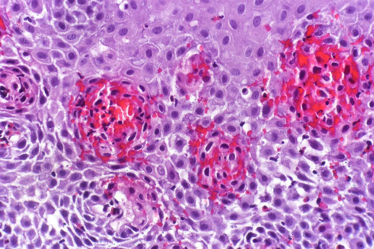
Identifying esophagitis and pinning down its exact subtype relies on a combination of imaging, direct visualization, and tissue analysis. An upper endoscopy allows the physician to inspect the esophageal wall with a camera while simultaneously snipping a small biopsy for histological confirmation of inflammation. A barium swallow—sometimes called an upper GI series or esophagram—coats the tract with a radiopaque contrast so that fluoroscopy and X-ray can reveal narrowing, ulceration, or structural irregularity. Laboratory work on the biopsy specimen can distinguish a fungal, viral, or bacterial pathogen, and a white-blood-cell scan can flag the eosinophilic infiltration characteristic of eosinophilic esophagitis. Clinically, the condition is sorted into several categories. Reflux esophagitis stems from acid backflow, with some research pointing toward cytokine-mediated pathways. Infectious forms are subdivided into fungal (notably Candida), viral (herpes simplex and cytomegalovirus), and rarer parasitic or bacterial variants, clustering around patients with weakened immunity. Drug-induced esophagitis results from medication-related tissue damage. Eosinophilic esophagitis, driven by allergen-triggered immune cells, and the even rarer lymphocytic esophagitis, first described in 2006, round out the taxonomy.
Complications and the Risk of Silent Progression
Left to its own devices, esophagitis follows a predictable and troubling trajectory. The persistent irritation that defines the condition prevents the mucosa from completing its normal repair cycle, and the resulting scar tissue progressively stiffens and narrows the tube. What begins as a mild burning sensation in the lower chest—worsened by lying flat or straining—can escalate to painful, obstructed swallowing, recurrent nausea, vomiting, abdominal cramping, and a persistent cough. If the inflammatory insult continues unchecked, discrete esophageal ulcers may form, adding a layer of localized tissue destruction to the diffuse swelling. More alarmingly, chronic reflux-driven inflammation can remodel the squamous lining into columnar tissue, a metaplastic change known as Barrett's esophagus, which carries a well-documented elevation in the risk of esophageal cancer. It is worth noting that the disease does not always announce itself; a meaningful subset of patients remain entirely asymptomatic until endoscopic or imaging workup reveals the damage. This silent progression underscores why early recognition and treatment of the underlying irritant—whether acid, drug, infection, or immune dysregulation—matter so much in preventing the cascade from inflammation to ulceration to malignancy.
Gallery
Frequently Asked Questions
Who is Esophagitis?
Esophagitis is a condition in which the mucosal lining of the esophagus — the muscular tube linking the pharynx to the stomach — becomes inflamed. It belongs to the gastroenterology field and is most frequently triggered by gastroesophageal reflux disease.
What are Esophagitis's signature moves?
Its typical 'attacks' include heartburn, difficulty swallowing (dysphagia), nausea, vomiting, abdominal pain, and a persistent cough, though some episodes remain entirely asymptomatic. The severity can swing from a mild, unnoticed irritation to a painful, debilitating flare.
What's Esophagitis's origin story?
The primary 'villain' behind most Esophagitis cases is GERD, where stomach acid repeatedly splashes back up and irritates the esophageal mucosa. While other triggers exist, chronic acid reflux is the dominant driver in the canon.
How do fans confirm Esophagitis in the field?
Clinicians typically lock in the diagnosis with an upper endoscopy paired with a tissue biopsy, often supplemented by an upper GI series or targeted laboratory tests. These tools help separate true esophageal inflammation from other sources of throat or chest discomfort.
Why is Esophagitis important to the overall canon?
Because GERD is so widespread, Esophagitis touches a large share of the population and acts as a key warning sign of ongoing acid-related tissue damage. If left unmanaged, the chronic inflammation it produces can progress to more serious esophageal complications, making early recognition and treatment critical.
More in Inflammatory diseases of the digestive system 1-22
Spotted an error? Know more?
Reader corrections go straight into our review queue. Suggest an edit · How this site is sourced