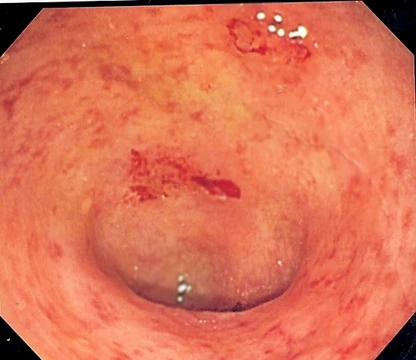
Duodenal lymphocytosis
A histological marker of duodenal inflammation often linked to coeliac disease.

Duodenal lymphocytosis, also known as lymphocytic duodenitis, lymphocytic duodenosis, or duodenal intraepithelial lymphocytosis, is a condition characterized by an increased number of intra-epithelial lymphocytes in biopsies of the duodenal mucosa when examined microscopically. It is often a feature of coeliac disease but may also be found in other disorders, making it a significant histological finding in gastroenterology.
Quick Facts
- Field
- Gastroenterology
- Causes
- Coeliac disease, environmental enteropathy and others
- Diagnosis
- Histological examination of duodenal biopsy
- Frequency
- 3–7% of people having duodenal biopsy
Facts from the source article.
Lore & Background
Duodenal lymphocytosis is diagnosed by accurate counting of intraepithelial lymphocytes during histological examination, with the requirement that the duodenal mucosa otherwise appears normal, specifically with preserved villous architecture. The condition is the least severe change in the Marsh classification for coeliac disease, known as Marsh I, and can progress to more severe stages involving villous atrophy and crypt hyperplasia.
Reader's Guide
The significance of duodenal lymphocytosis lies in its role as a sensitive but non-specific histological finding. While it is a hallmark of coeliac disease, particularly in untreated or partially treated cases, it can also arise from a wide range of causes including environmental enteropathy, autoimmune enteropathy, small intestinal bacterial overgrowth, NSAID damage, Helicobacter pylori infection, other infections, and Crohn's disease. Diagnosis requires careful exclusion of other causes and may involve serological testing for coeliac-associated antibodies and HLA-DQ2/8 genotyping. Management focuses on treating the underlying disorder, such as a gluten-free diet for coeliac disease or antimicrobial therapy for infections. The condition's prognosis varies; in cases linked to coeliac disease, a gluten-free diet can reduce intraepithelial lymphocyte numbers, and progression to overt coeliac disease occurred in 23 of 85 patients over two years in one series, particularly in those with diarrhea, thyroiditis, weakness, or folate deficiency.
Did You Know?
- Intra-epithelial lymphocytes are normally present in the intestine, with greater numbers in the crypts and jejunum, and are distinct from those in the lamina propria.
- Around 33% of duodenal lymphocytosis cases have the HLA-DQ2 allele, which is found in over 90% of people with coeliac disease.
- Helicobacter pylori infection is a recognized cause of duodenal lymphocytosis in both children and adults.
- In one series, a subset of patients with duodenal lymphocytosis later developed gluten sensitivity and coeliac disease, though exact figures vary across studies.
Microscopic Identity and Prevalence
Duodenal lymphocytosis—also referred to in the literature as lymphocytic duodenitis, lymphocytic duodenosis, or duodenal intraepithelial lymphocytosis—describes a histological finding rather than a single disease entity. When a pathologist examines a duodenal mucosal biopsy under the microscope, the hallmark is an elevated density of intra-epithelial lymphocytes (IELs) residing within the epithelial layer of the duodenum. The conventional threshold for calling this abnormal sits at roughly 20 to 25 IELs per 100 enterocytes. These lymphocytes, predominantly T cells, are a normal resident population of the intestinal epithelium, with baseline numbers tending to be higher in the crypt regions and in the jejunum compared with the duodenum. They are anatomically and functionally distinct from the lymphocytes that populate the lamina propria beneath the epithelium. In routine practice, an IEL count above the threshold is encountered in approximately three percent of duodenal biopsies, though some series report figures climbing toward seven percent, suggesting the finding may be increasingly recognized as case mixes shift and counting techniques refine.
A Broad Differential: Beyond Coeliac Disease
Although duodenal lymphocytosis is most frequently discussed in the context of coeliac disease, the underlying aetiology is far from singular. The differential spans a wide spectrum of conditions. Tropical sprue and other forms of environmental enteropathy can provoke the same epithelial lymphocyte infiltration. Autoimmune enteropathy, small intestinal bacterial overgrowth, and mucosal injury from non-steroidal anti-inflammatory drugs all feature on the list. Infectious agents play a notable role: Helicobacter pylori colonisation, Cryptosporidiosis, and Giardiasis have each been linked to elevated IEL counts. Crohn's disease, when it involves the duodenum, may also present with this histological pattern. In paediatric populations, Helicobacter infection is a common endoscopic finding, and while IEL counts tend to be marginally higher in its presence, clinicians have generally not regarded it as a meaningful driver of the lymphocytosis in children. The breadth of this differential underscores that an elevated IEL count is a shared histological response to diverse mucosal insults rather than a pathognomonic marker of any one disease.
Unraveling the Diagnosis: The Coeliac Connection
Confirming duodenal lymphocytosis requires meticulous counting of IELs during histological examination, with the critical stipulation that the surrounding duodenal architecture—villous structure in particular—remains normal. This distinguishes the finding from the more advanced histological changes seen in later Marsh stages of coeliac disease, where villous atrophy and crypt hyperplasia accompany the lymphocytic infiltration. In untreated or partially treated coeliac disease, isolated duodenal lymphocytosis corresponds to Marsh stage I, the mildest category in that classification. Serological testing adds diagnostic weight: IgA endomysial antibodies and anti-tissue transglutaminase antibodies, both highly sensitive and specific for coeliac disease, were positive in roughly eleven percent of duodenal lymphocytosis cases, implying a definite coeliac aetiology in that subset. HLA typing provides further context; the HLA-DQ2 allele, present in over ninety percent of coeliac patients, was detected in about one-third of lymphocytosis cases, while the absence of both HLA-DQ2 and the rarer HLA-DQ8 makes coeliac disease highly unlikely. In antibody-negative individuals, a gluten challenge followed by symptomatic response to a gluten-free diet has been employed to firm up the diagnosis, yielding a confirmed coeliac label in approximately twenty-two percent of a series exceeding two hundred adult patients.
Treatment Pathways and Long-Term Outlook
Because duodenal lymphocytosis is a histological descriptor rather than a standalone disease, management is directed at whatever underlying condition is identified. In cases where coeliac disease is confirmed, a strict gluten-free diet remains the cornerstone of therapy, and this intervention has been shown to reduce IEL numbers over time. When an infection such as Helicobacter pylori, Cryptosporidium, or Giardia is the culprit, targeted antimicrobial or antiparasitic treatment addresses the root cause. NSAID-related mucosal injury, by contrast, calls for withdrawal or substitution of the offending agent. The long-term prognosis hinges on whether the lymphocytosis is an isolated finding or a harbinger of evolving gluten sensitivity. In one prospective series of eighty-five patients, twenty-three progressed to overt coeliac disease within two years. Several clinical and serological features flagged those at higher risk: positive coeliac antibodies, the presence of HLA-DQ2 or HLA-DQ8, a family history of the condition, and accompanying symptoms such as chronic diarrhoea, thyroiditis, unexplained weakness, and folate deficiency. Recognising these predictors allows clinicians to monitor more closely and intervene early, potentially preventing the full histological and nutritional consequences of established coeliac disease.
Frequently Asked Questions
What exactly is Duodenal lymphocytosis?
It is a histological finding in which duodenal mucosal biopsies reveal an elevated count of intra-epithelial lymphocytes, typically exceeding 20 to 25 per 100 enterocytes. The condition is also referred to as lymphocytic duodenitis, lymphocytic duodenosis, or duodenal intraepithelial lymphocytosis.
How common is Duodenal lymphocytosis in routine duodenal biopsies?
It is identified in roughly 3% of all duodenal biopsies, with some case series reporting figures as high as 7%. This makes it a notable but not uncommon microscopic observation in gastroenterology practice.
What is the connection between Duodenal lymphocytosis and coeliac disease?
It is a recognized feature of untreated or partially treated coeliac disease, corresponding to Marsh stage I mucosal changes. In one series tracking over 200 adult patients, approximately 22% ultimately received a confirmed coeliac diagnosis.
Does Duodenal lymphocytosis always indicate coeliac disease?
No, it can be present in a range of other gastrointestinal disorders as well. Only about 11% of affected individuals test positive for IgA endomysial or anti-transglutaminase antibodies, and roughly one-third carry the HLA-DQ2 allele, so the finding alone is not diagnostic.
Why is Duodenal lymphocytosis considered important in gastroenterology?
It acts as a histological red flag for duodenal inflammation that prompts clinicians to pursue additional serological and genetic testing, particularly for coeliac disease. Because it overlaps with multiple conditions, it is treated as one component of a broader diagnostic workup rather than a standalone verdict.
More in Inflammatory diseases of the digestive system 1-22
Spotted an error? Know more?
Reader corrections go straight into our review queue. Suggest an edit · How this site is sourced