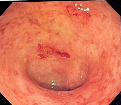
Epiploic appendagitis
Self-limiting colon fat inflammation mimicking surgical abdomen.

Hellerhoff · CC BY-SA 3.0
Epiploic appendagitis (EA) is an uncommon, benign, self-limiting inflammatory process of the epiploic appendices—small, fat-filled sacs along the surface of the colon and rectum. It is significant as a condition that mimics more serious abdominal emergencies such as acute appendicitis, diverticulitis, and cholecystitis, and is increasingly diagnosed incidentally on CT scans.
- Field
- Gastroenterology / Radiology
- Known for
- Self-limiting inflammation of epiploic appendices, often misdiagnosed as diverticulitis or appendicitis
- Typical age range
- 12 to 82 years, most common in 40s and 50s
- Sex predominance
- Men slightly more affected than women
- Common misdiagnosis
- Diverticulitis (left-sided pain) or appendicitis (right-sided pain)
Lore & Background
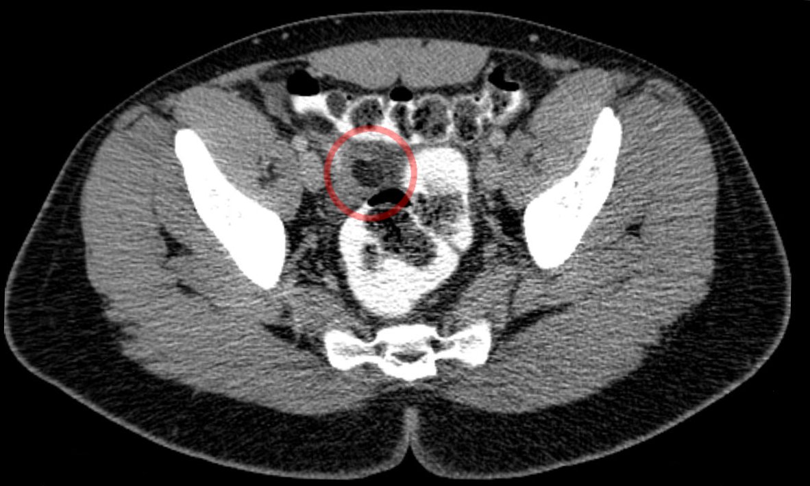
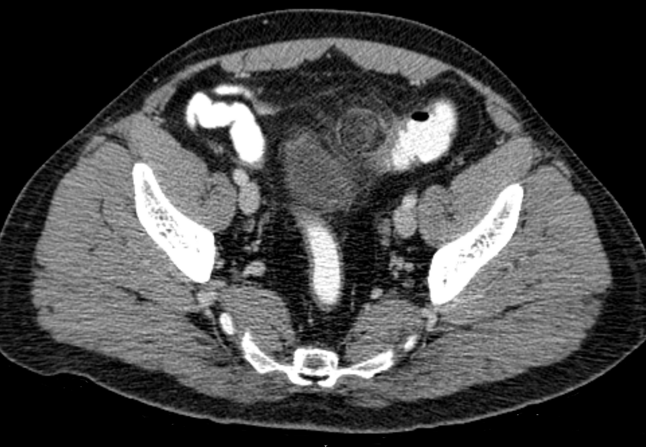
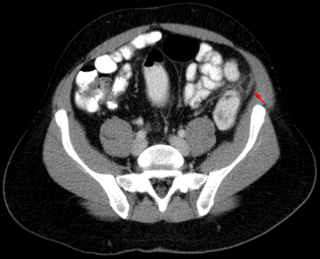
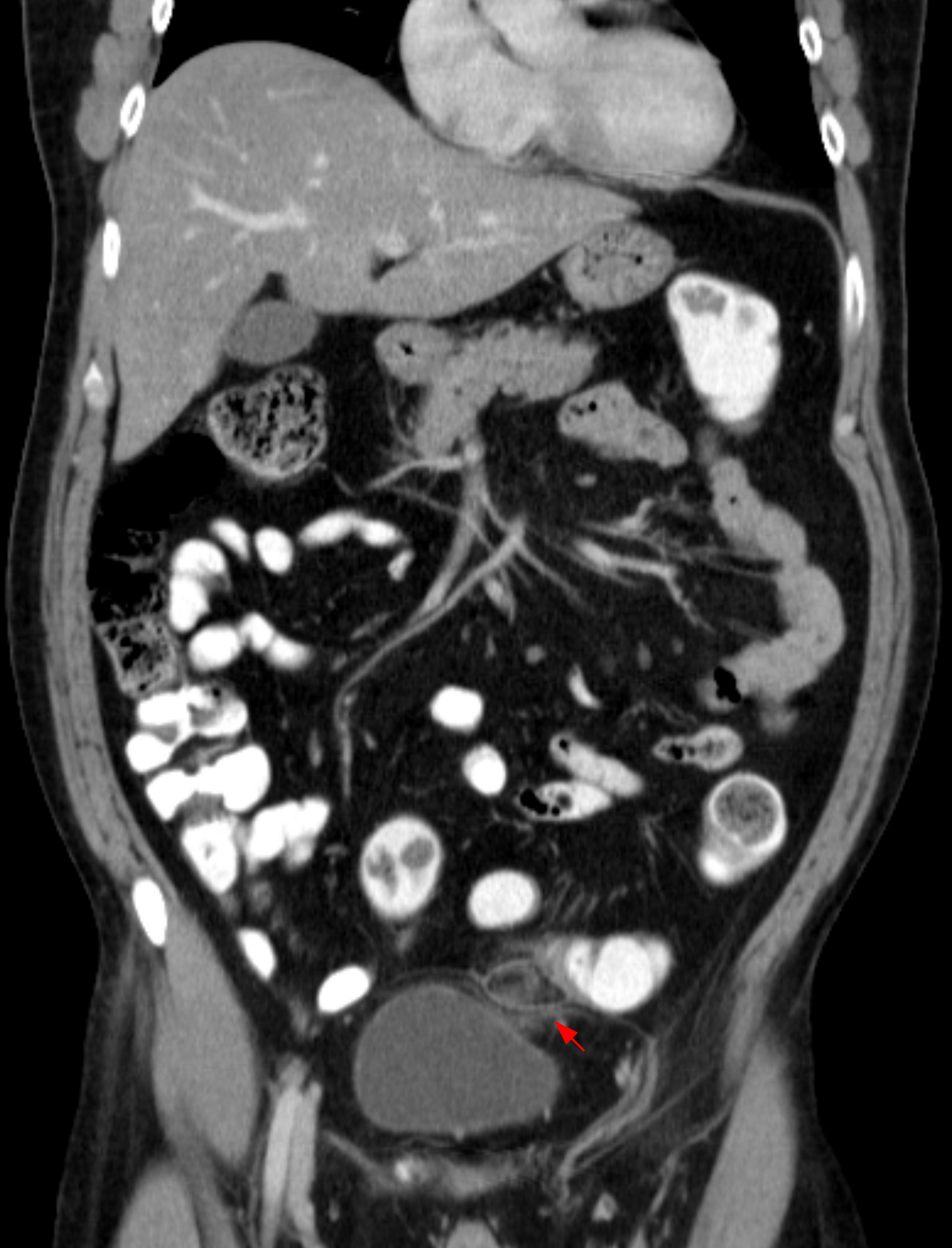
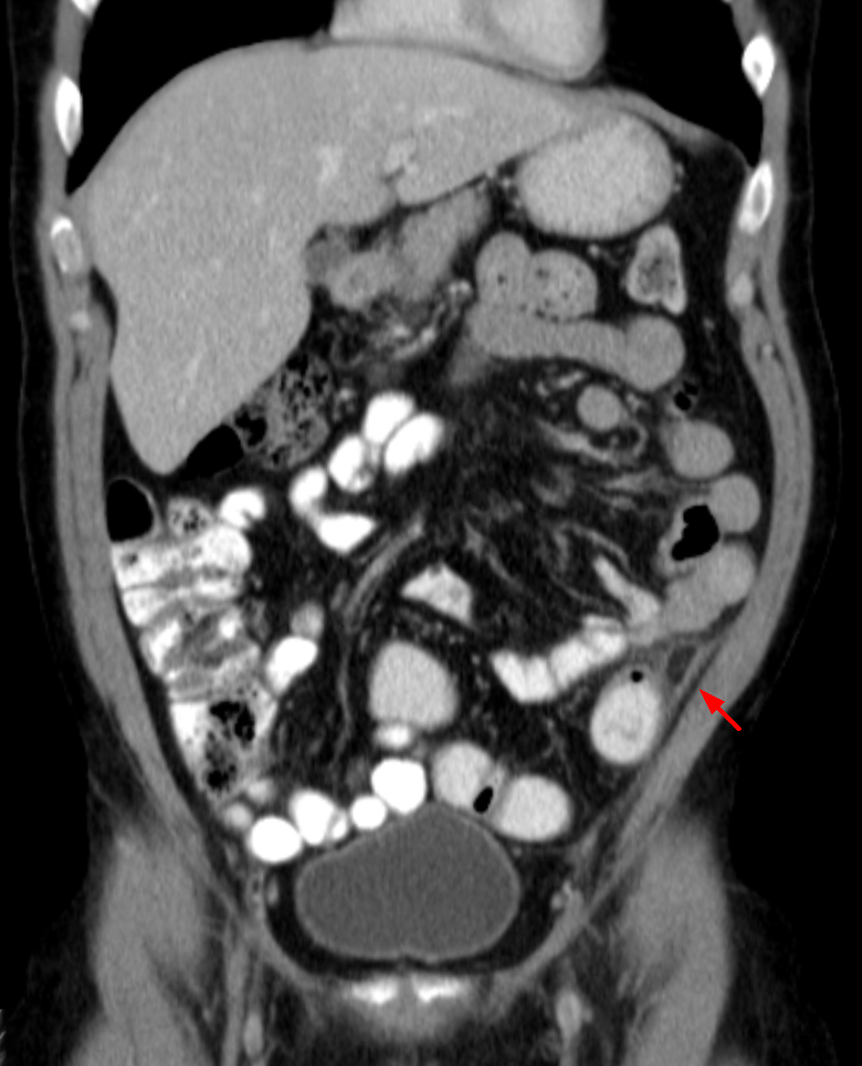
Epiploic appendagitis results from torsion or venous thrombosis of the epiploic appendages, causing sharp or stabbing abdominal pain often worsened by defecation or urination. The condition is usually diagnosed incidentally on CT scan, which shows characteristic oval fat-density lesions surrounded by inflammation. Initial lab studies are typically normal, and patients do not usually have fever, vomiting, or leukocytosis, helping distinguish it from diverticulitis.
While self-limiting, epiploic appendagitis can cause severe pain. Most sources recommend conservative management with anti-inflammatory medications and analgesics, with surgery not recommended in nearly all cases. However, Sand and colleagues recommend laparoscopic excision to prevent recurrence. Rare complications include peritoneal loose bodies, which can cause urinary retention or bowel obstruction.
The condition is more common in patients over 40, with reported ages from 12 to 82 years. It is associated with obesity, hernia, and unaccustomed exercise. Symptoms typically resolve within two weeks under non-invasive treatment, and hospitalization is not necessary.
Reader's Guide
Epiploic appendagitis holds significance as a diagnostic pitfall in acute abdominal pain. Its symptoms closely mimic those of surgical emergencies such as appendicitis, diverticulitis, and cholecystitis, yet it requires no surgical intervention. The increasing use of abdominal CT for evaluating lower abdominal pain has led to more frequent diagnosis of this self-limiting condition, sparing patients unnecessary surgery. The condition's epidemiology—predominantly in men in their 40s and 50s, associated with obesity and unaccustomed exercise—helps clinicians consider it in the differential. The article notes a dispute regarding management: while most sources advise conservative treatment with NSAIDs and analgesics, Sand and colleagues recommend laparoscopic excision to prevent recurrence. This unresolved disagreement reflects ongoing clinical debate. The condition's legacy lies in its role as a benign mimic of serious disease, emphasizing the value of imaging in modern abdominal diagnosis.
Did You Know?
- Epiploic appendagitis is caused by torsion or venous thrombosis of fat-filled sacs along the colon.
- Pain is characteristically intense during or after defecation or urination due to traction on the lesion.
- The condition is usually diagnosed incidentally on CT scan performed to exclude more serious conditions.
- Rarely, epiploic appendagitis can result in a peritoneal loose body that may cause urinary retention or bowel obstruction.
Anatomy & Pathophysiology
The epiploic appendages—also known as appendices epiploicae—are small, fat-filled projections that line the outer surface of the upper and lower colon as well as the rectum. A typical colon carries roughly fifty to one hundred of these structures, arranged in two parallel rows (anterior and posterior) that run alongside the superficial layer of the taenia coli. Each appendage measures between half a centimetre and five centimetres in length and is tethered to the colonic wall by a short vascular stalk containing one or two small arterioles and a venule. When one of these stalks undergoes torsion—a twisting or wrenching motion—or when the venous drainage becomes obstructed by a thrombus, the appendage loses adequate blood supply and becomes acutely inflamed. This ischemic insult triggers a localized inflammatory cascade that, although entirely benign and self-limiting, produces a strikingly painful episode. The process is sometimes still referred to by older labels such as appendicitis epiploica or simply appendagitis, though clinicians have largely abandoned those terms to sidestep confusion with true acute appendicitis.
Clinical Presentation & Diagnostic Challenge
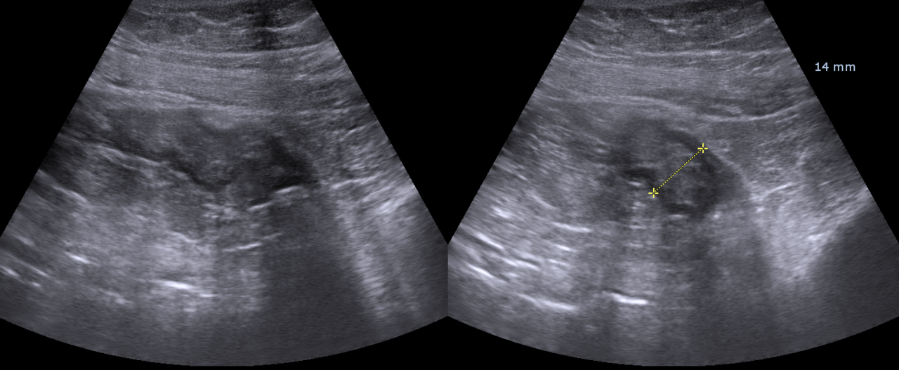
Patients with epiploic appendagitis typically present with a sudden, localized, sharp or stabbing pain in the lower abdominal region—most often the left lower quadrant, though right-sided or central presentations also occur. The discomfort is characteristically aggravated by straining during defecation or urination, particularly with sigmoid-type lesions, because the mechanical traction on the inflamed pedicle intensifies the pain. Nausea and occasional vomiting may accompany the episode, but fever, leukocytosis, and changes in bowel habit are generally absent. Because the symptom profile overlaps so closely with diverticulitis, acute appendicitis, and even cholecylitis, the condition is misdiagnosed in the majority of cases. Initial laboratory work is usually unremarkable, and the diagnosis is frequently made only incidentally when a CT scan—ordered to rule out more dangerous pathology—reveals the inflamed appendage. Ultrasound can also confirm the finding. Distinguishing EA from omental infarction or true diverticulitis on imaging can be particularly difficult, since diverticulitis may secondarily inflame the appendages and omental infarction lacks the bowel-wall thickening seen in diverticular disease.
Treatment & Management
Despite the intensity of the pain it produces, epiploic appendagitis is fundamentally a self-resolving condition, and the prevailing clinical consensus is to manage it conservatively. The standard approach involves a course of anti-inflammatory medication combined with a moderate-to-severe analgesic, tailored to the severity of the individual's symptoms. In nearly all reported cases, surgical intervention is neither necessary nor recommended, since the inflammation resolves on its own without leaving lasting structural damage. However, a notable dissenting view has been put forward by Sand and colleagues, who advocate for laparoscopic excision of the affected appendage in most cases. Their rationale centres on preventing recurrence: by removing the inflamed structure, they argue, the patient is spared the risk of a repeat episode. This surgical stance remains a minority position, and the broader medical community continues to favour watchful waiting and pharmacologic symptom control as the first-line strategy for this benign, self-limiting process.
Rare Complications & Demographics
Although epiploic appendagitis is overwhelmingly benign, a rare but documented sequela is the formation of a peritoneal loose body. This occurs when a torsed, infarcted, or fully detached appendage undergoes progressive fibrosis—essentially turning into a free-floating mass of dead fibrous tissue encased in multiple layers of calcium-salt deposits. If such a loose body grows sufficiently large, it can impinge on adjacent structures, producing urinary retention or even a bowel obstruction. In terms of who is affected, the condition shows a clear demographic pattern: it is most frequently encountered in patients in their fourth and fifth decades of life, with a slight predominance in men. Nevertheless, case reports span an age range from twelve to eighty-two years, confirming that no single age group is immune. The condition is uncommon enough that many clinicians encounter it only rarely in their careers, which contributes to the high rate of initial misdiagnosis and the reliance on cross-sectional imaging for definitive identification.
Gallery
Frequently Asked Questions
Who is Epiploic appendagitis?
Epiploic appendagitis (EA) is a benign, self-resolving inflammatory episode that targets the tiny fat-filled sacs (epiploic appendices) draped along the outer surface of the colon and rectum. It sits at the intersection of gastroenterology and radiology and is classified as uncommon rather than routine.
What are Epiploic appendagitis's powers/role?
Its signature 'power' is convincingly imitating far more dangerous abdominal emergencies—left-sided cases get mistaken for diverticulitis, while right-sided ones are confused with acute appendicitis or even cholecystitis. This mimicry is what keeps it a persistent diagnostic trap in emergency imaging.
Why is Epiploic appendagitis important?
It matters because recognizing it prevents unnecessary laparotomies and antibiotic courses that would otherwise be directed at a harmless fat-sac irritation. Its rising detection rate on cross-sectional CT scans has made it one of the most frequently 'uncovered' incidental findings in modern abdominal imaging.
Who does Epiploic appendagitis typically target?
The condition can strike anywhere from age 12 to 82, with peak incidence in the 40s and 50s, and shows a slight male predominance over female cases. No single risk factor defines a 'susceptible' patient, so it tends to appear as an unexpected plot twist in an otherwise unremarkable abdominal workup.
More in Inflammatory diseases of the digestive system 1-22
Spotted an error? Know more?
Reader corrections go straight into our review queue. Suggest an edit · How this site is sourced