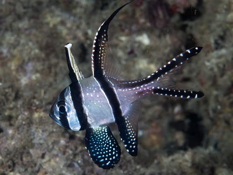
Lateral stripe
One band of pigment that tells a reef's residents exactly who you are.

A lateral stripe is a longitudinal color band running along the flank of a marine fish, typically extending from the operculum or just behind the gill cover toward the caudal peduncle. It is one of the most widespread and immediately recognizable markings across reef and pelagic ichthyofauna, appearing in damselfishes (Pomacentridae), wrasses (Labridae), anthias (Anthiidae), gobies (Gobiidae), and numerous other families. Unlike the lateral line—a sensory canal embedded within the scale plates that detects water pressure and vibration—a lateral stripe is a pigment-based visual pattern produced by chromatophores and iridophores in the dermal layer.
In the field, a lateral stripe functions as a multi-purpose signal: it can break up the body outline against the dappled light of a coral thicket, serve as a species-recognition cue during territorial or courtship displays, and in some taxa act as a deflection mark that draws a predator's strike away from the head toward the less vulnerable flank. Its width, color, and exact starting point vary enough between closely related species that experienced ichthyologists use it as a primary field key.
- Type
- Morphological color pattern (descriptive term, not a taxon)
- Typical location
- Mid-lateral flank, operculum to caudal peduncle
- Pigment source
- Dermal chromatophores and iridophores
- Distinct from
- Lateral line (neurosensory canal in scales)
- Ecological roles
- Camouflage, species recognition, predator deflection
- Families in which commonly found
- Pomacentridae, Labridae, Anthiidae, Gobiidae, Acanthuridae, and others
Lore & Background
In reef ichthyology the lateral stripe occupies a peculiar niche: it is not a taxonomic unit, yet it is often the single feature that separates two otherwise nearly identical species. Two damselfish of the same genus may differ only in whether the stripe begins at the eye or behind the operculum, or whether it is a clean black line versus a broad yellowish band edged in blue. Field guides and online databases routinely list stripe position, width, and color as diagnostic characters precisely because the rest of the body plan is so conserved within a genus.
The stripe's biological origin is straightforward. Melanophores, xanthophores, and iridophores in the dermis lay down pigment in a narrow longitudinal row during embryonic development, a pattern that is genetically canalized and expressed with little individual variation. In species that change color with sex, social status, or time of day, the stripe may broaden, fade, or shift hue, but its position along the flank remains stable. This stability is what makes it a reliable recognition signal: a conspecific can identify a rival or a mate at a glance, even in the turbid, low-contrast light of a shaded reef overhang.
Predator ecology adds another layer. Several studies on damselfish and wrasse behavior note that a bold lateral stripe can cause a striking predator—often a larger wrasse or grouper—to misjudge the fish's head position and bite the flank instead. The stripe effectively creates a visual 'false head' at the posterior end of the band. Whether this deflection hypothesis fully explains the stripe's prevalence remains debated, but it is one of the most cited selective pressures in the literature on reef-fish coloration.
Reader's Guide
What the eye catches first is the stripe itself: a single, unbroken longitudinal band of dark pigment—black, deep brown, or in some species a saturated yellow or blue—running along the mid-lateral flank. Unlike the vertical bars seen on many damselfish or the reticulated patterns of some wrasses, the lateral stripe is a clean, parallel-edged line that does not break across the body. Trace it from its anterior origin: in some species it begins at the posterior margin of the operculum, in others it starts just behind the eye, and in a few it extends forward to the snout tip. Follow it posteriorly and note whether it tapers to a point at the caudal peduncle, broadens into a saddle across the dorsal base, or continues unbroken onto the caudal fin.
Next, examine the edges. A sharply demarcated stripe with a hard boundary against the ground color suggests a high-contrast melanophore band; a softer, feathered edge indicates a gradient of xanthophores or iridophores. In some species the stripe is bordered by a thin white or pale blue line, adding a second visual element that increases contrast against the surrounding water.
Look at the scales within the stripe. In many pomacentrids the scales over the stripe are the same size and shape as those on the flanks, but the pigment sits in the dermis beneath them, so the scale outline remains visible through the color. In a few labrids the scales within the stripe are slightly enlarged or have a different micro-ornamentation, giving the band a subtly different texture under raking light.
Finally, compare the stripe's width to the interocular distance. A stripe narrower than the eye diameter reads as a 'line'; one wider reads as a 'band.' This proportional relationship, combined with the exact anterior and posterior endpoints, is often the single most reliable field character for separating congeners that differ in no other visible respect.
More in Description
Elsewhere in Banggai cardinalfish
Spotted an error? Know more?
Reader corrections go straight into our review queue. Suggest an edit · How this site is sourced