Vitiligo
Chronic autoimmune disorder causing skin depigmentation.

DataBase Center for Life Science (DBCLS) · CC BY 4.0
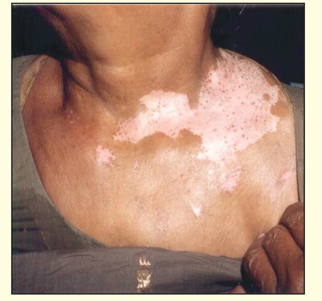
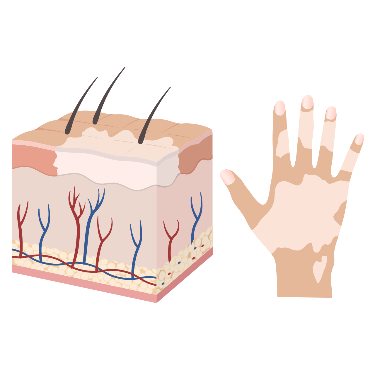
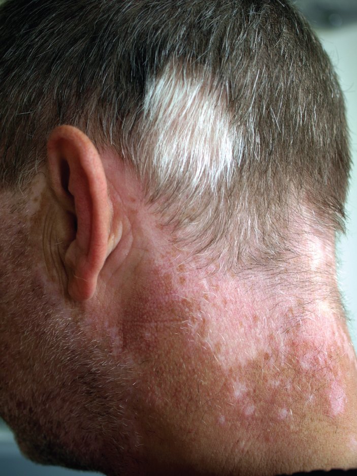
Vitiligo is a chronic autoimmune disorder that causes the skin to lose pigment in patches that vary in size and can appear anywhere on the body. The condition is linked to aberrant attachments between melanocytes and laminins, and is thought to be caused by immune system changes with potential genetic factors.
- Field
- Dermatology / Autoimmune disorder
- Known for
- Chronic depigmentation of the skin
- Symptoms
- Pale, patchy areas of depigmented skin, often on extremities
- Common treatments
- Topical immunosuppressants, ultraviolet light therapy (311-nm UVB) with antioxidants
Lore & Background
Vitiligo often first appears by young adulthood and may be triggered by environmental factors, sun or chemical exposure, stress, and physical trauma. The most common form, nonsegmental vitiligo, tends to affect more skin over time. In antiquity, the disorder was often conflated with leprosy. Some public figures have had vitiligo, such as the singer Michael Jackson, who publicly acknowledged his condition in 1993.
Reader's Guide
Vitiligo is significant as a chronic condition that affects pigmentation and can lead to stigmatization, depression, and similar mood disorders. Its development involves immune system attacks on melanocytes, with genetic susceptibilities and environmental triggers. The disorder also appears in other mammals, such as Arabian horses. Treatments include topical immunosuppressants and ultraviolet light therapy, though segmental vitiligo is highly resistant to medical treatment. The condition has been linked to autoimmune and inflammatory diseases including Hashimoto's thyroiditis, scleroderma, rheumatoid arthritis, type 1 diabetes, psoriasis, Addison's disease, pernicious anemia, alopecia areata, systemic lupus erythematosus, and celiac disease.
A Place Among Many
Vitiligo occupies one thread within a broad family of conditions collectively termed leucism, a label rooted in the Greek word leukós, meaning white. It shares this classification with piebaldism, Waardenburg syndrome, Chédiak–Higashi syndrome, flavism, isabellinism, xanthochromism, axanthism, amelanism, and mutations involving melanophilin. What unites all of these is a partial loss of pigmentation that produces white, pale, or patchy coloration across skin, hair, feathers, scales, or cuticles. Crucially, the eyes remain unaffected in every one of these conditions. Vitiligo is therefore not a standalone anomaly but one expression within a wider spectrum of pigment-related genetic conditions, each producing its own distinctive pattern of depigmentation while sharing the common thread of reduced or absent pigment cell function.
The Neural Crest Story
At its developmental root, vitiligo and its leucistic relatives trace back to a critical window in embryonic life. Pigment cells originate as multipotent precursors that must differentiate and migrate from the neural crest into target tissues such as skin, hair follicles, and feather structures. When this process falters—whether in the differentiation step or the migratory journey—the result is a body surface lacking the cells capable of producing pigment. Because every pigment cell type springs from the same precursor, a disruption here can diminish all pigment varieties simultaneously. This is a fundamentally different mechanism from albinism, where the melanocyte is present and structurally intact but simply fails to manufacture melanin. In vitiligo's case, the problem lies upstream: the pigment cells never fully develop or never arrive at their destination in the first place.
Not Albinism: A Critical Distinction
One of the most persistent misconceptions surrounding vitiligo is its conflation with albinism, yet the two conditions operate through entirely different biological logic. Albinism involves a melanocyte that exists but cannot produce melanin, leaving the animal with a pale yellow tint in species that also carry other pigment cells such as xanthophores. Vitiligo, as a leucistic condition, reflects an absence or severe reduction of pigment cells altogether. This distinction carries visible consequences for the eyes: albinism can affect the retinal pigmented epithelium and iris, sometimes revealing pink pupils through underlying blood vessels. Leucistic conditions, including vitiligo, spare the eyes because the melanocytes of the retinal pigmented epithelium arise from an independent developmental origin—an out-pouching of the neural tube that forms the optic cup and retina—rather than from the neural crest. Thus, vitiligo-affected individuals typically retain normally colored eyes.
Genes, Patches, and Unpredictable Patterns
The genetic architecture behind vitiligo and related leucistic conditions involves mutations in genes such as c-kit, mitf, and EDNRB. Rather than producing a uniform, all-white appearance, these conditions more commonly manifest as localized or incomplete hypopigmentation—irregular patches of white set against an otherwise normally colored and patterned body. This pied or piebald effect is the more frequent presentation. What makes the pattern particularly striking is its unpredictability: the ratio of white to normally pigmented tissue can shift dramatically from one generation to the next, vary between siblings born to the same parents, and even differ among members of a single litter. This variability means that no two affected individuals present identically, making vitiligo a condition of remarkable individual expression rather than a single fixed phenotype.
Gallery
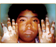
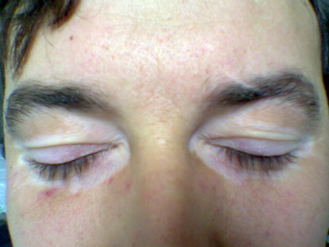
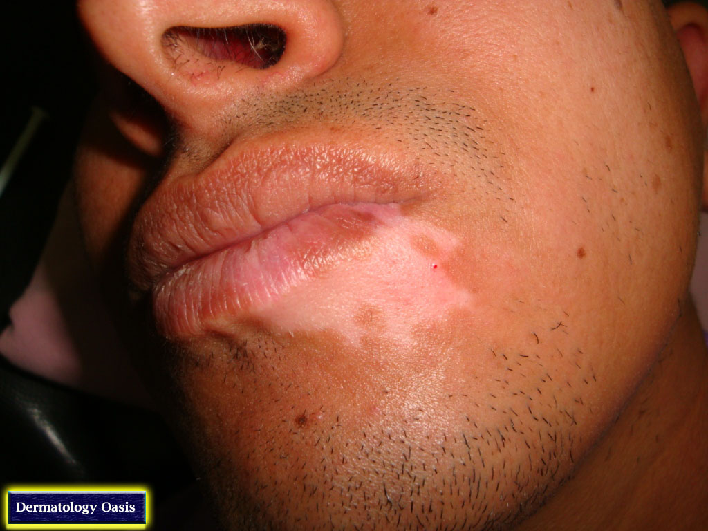
Frequently Asked Questions
Who is Vitiligo?
Vitiligo is a long-term autoimmune condition classified under dermatology, best known for its signature effect of stripping pigment from localized patches of skin. It is not contagious and does not damage internal organs, but it permanently alters the visible appearance of affected areas.
What are Vitiligo's powers or role?
Its defining 'ability' is the progressive loss of melanin in specific skin regions, producing pale, irregular patches most often on the hands, feet, face, and around body openings. Mechanistically, it disrupts the structural bond between pigment-producing melanocytes and laminin proteins in the dermis.
What causes Vitiligo to appear?
The precise trigger is still debated, but the leading explanation involves a misdirected immune response that attacks melanocytes, layered on top of inherited genetic susceptibility. Abnormal attachment between melanocytes and laminins is thought to be a key structural step in the depigmentation process.
How do people treat or manage Vitiligo?
Standard management includes topical immunosuppressant creams to quiet the overactive immune attack and targeted 311-nanometer UVB phototherapy paired with antioxidant support. Because responses vary widely, regimens are usually tailored to each individual's pattern and severity.
More in Injuries And Conditions 1-24
Spotted an error? Know more?
Reader corrections go straight into our review queue. Suggest an edit · How this site is sourced