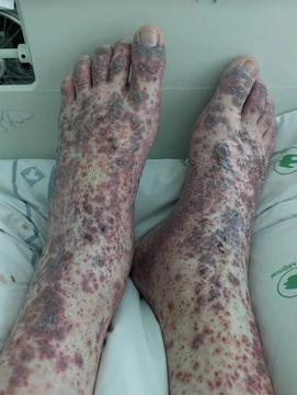
Vasculitis
A group of disorders destroying blood vessels through inflammation.

Vasculitis is a group of disorders characterized by inflammation that destroys blood vessels, affecting both arteries and veins. Lymphangitis, inflammation of lymphatic vessels, is sometimes considered a type of vasculitis. The condition is primarily caused by leukocyte migration and resultant damage, though inflammation of veins (phlebitis) or arteries (arteritis) alone are separate entities.
Quick Facts
- Field
- Rheumatology, Immunology
- Pronounce
- v · æ · s · k · j · ʊ · ˈ · l · aɪ · t · ɪ · s
- Symptoms
- Weight loss, fever, myalgia, purpura, abdominal pain
- Complications
- Gangrene, Myocardial infarction
Facts from the source article.
Lore & Background
Vasculitis encompasses a range of conditions that damage blood vessels via inflammation, driven primarily by leukocyte migration. The clinical presentation depends largely on the diameter of the affected vessels, with non-specific symptoms such as fever, headache, fatigue, myalgia, weight loss, and arthralgia being common. Skin manifestations, including purpura, nodules, livedo reticularis, skin ulcers, and purpuric urticaria, can occur in all forms, even large vessel vasculitides.
Reader's Guide
The 2012 Chapel Hill Consensus Conference provides the highest classification level, distinguishing primary systemic, secondary, and single-organ vasculitides. Primary systemic vasculitis is categorized by vessel size: large-vessel (e.g., Takayasu arteritis, giant cell arteritis), medium-vessel (e.g., polyarteritis nodosa, Kawasaki disease), small-vessel (including ANCA-associated and immune complex types), and variable-vessel (e.g., Behcet's disease, Cogan's syndrome). Secondary vasculitides are triggered by underlying conditions such as infections (e.g., hepatitis B and C, HIV), systemic illnesses, cancer, or drug exposure. Diagnosis involves laboratory tests showing inflammation (e.g., elevated ESR, CRP), biopsy of affected tissue, or imaging like angiogram or FDG-PET/CT for large vessel vasculitis. Treatment focuses on suppressing inflammation and the immune system, typically with corticosteroids like prednisone, and sometimes other immunosuppressants or antimicrobials if infection is present.
Did You Know?
- The 2012 Chapel Hill Consensus Conference classifies vasculitis into primary systemic, secondary, and single-organ types.
- Large vessel vasculitis includes Takayasu arteritis and giant cell arteritis, affecting the aorta and its major branches.
- Diagnosis can be confirmed by biopsy of involved tissue, showing the pattern of blood vessel inflammation.
The Inflammatory Mechanism Behind Vessel Destruction
Vasculitis encompasses a family of disorders united by one destructive process: the immune system's inflammatory assault on the walls of blood vessels. Unlike isolated phlebitis or arteritis, which target veins or arteries in isolation, vasculitis can strike both arterial and venous structures simultaneously, and in some classifications even extends to lymphatic vessels through a condition called lymphangitis. The central driver of this damage is the migration of leukocytes into the vessel wall, where their presence triggers progressive tissue injury. In certain presentations, this process takes the form of leukocytoclasis, in which nuclear debris shed by infiltrating neutrophils physically tears apart the vascular architecture. Clinically, this neutrophil-driven destruction most often surfaces as palpable purpura, a raised purpuric lesion that signals small-vessel compromise. Because the underlying mechanism is leukocyte-mediated rather than purely mechanical, the resulting damage tends to be chronic and multi-organ, making early recognition of the inflammatory cascade critical to limiting progressive vascular injury and preserving organ function.
The Chapel Hill Taxonomy: Sorting a Complex Family
The 2012 Chapel Hill Consensus Conference established the framework still used to organize vasculitis into manageable categories. At the broadest level, the system separates primary systemic vasculitis, secondary vasculitis triggered by an underlying condition or exposure, and single-organ vasculitis confined to one tissue such as the gut, skin, or peripheral nerves. Within primary systemic disease, the critical dividing line is vessel caliber. Large-vessel vasculitis, exemplified by Takayasu arteritis and giant cell arteritis, preferentially targets the aorta and its principal branches. Medium-vessel forms, including polyarteritis nodosa and Kawasaki disease, center on the arteries that irrigate internal organs. Small-vessel vasculitis splits into two mechanistic subgroups: ANCA-associated necrotizing vasculitis (encompassing granulomatosis with polyangiitis, eosinophilic granulomatosis with polyangiitis, and microscopic polyangiitis) and immune-complex-mediated types such as IgA vasculitis, cryoglobulinemic vasculitis, and anti-glomerular basement membrane disease. Finally, variable-vessel vasculitis, represented by Behcet's disease and Cogan's syndrome, defies size-based sorting because it can involve arteries, veins, and capillaries of every diameter without a clear predilection.
Reading the Clinical Clues: Symptoms and Diagnostic Pathways
Because vasculitis can mimic dozens of other conditions, its early presentation is often nonspecific. Patients frequently report fever, persistent headache, profound fatigue, muscle aches, unexplained weight loss, and joint pain—symptoms that could point to virtually any inflammatory or infectious process. The skin, however, often provides the most visible clue: purpura, subcutaneous nodules, livedo reticularis, skin ulcers, and purpuric urticaria can appear across all vessel sizes, even in large-vessel disease. Laboratory workup in active cases typically reveals elevated erythrocyte sedimentation rate and C-reactive protein, anemia, a raised white blood cell count with possible eosinophilia, and sometimes detectable ANCA titers or hematuria. Definitive confirmation, though, rests on tissue biopsy of the affected organ—skin, sinus, lung, nerve, brain, or kidney—where the pathologist can directly visualize the pattern of vascular inflammation and the presence or absence of immune deposits. When biopsy is impractical, angiography can reveal characteristic inflammatory patterns in the vessel lumen, and brain SPECT imaging may document reduced cerebral perfusion in neurologically involved cases.
Beyond Autoimmunity: The Many Triggers of Vascular Inflammation
While dysregulated autoimmune responses represent the most prevalent family of vasculitis etiologies, the condition is far from exclusive to immune self-attack. Infections remain a significant secondary trigger: hepatitis B and C, HIV, infective endocarditis, and tuberculosis are all recognized as important causes, either by directly injuring the vessel wall or by provoking immune-mediated secondary events that spiral into a vasculitic syndrome. Even simple vascular thrombosis, which ordinarily affects only the lumen, can—through the gradual process of thrombus organization—evolve into a more chronic vasculitic picture. Systemic autoimmune diseases such as rheumatoid arthritis can give rise to rheumatoid vasculitis, and malignancy or certain drug exposures have also been implicated, though conclusively proven pathogenic links remain limited for many of these associations. Notably, with the exception of rheumatoid vasculitis, the majority of secondary vasculitis forms are exceedingly rare in clinical practice. This diversity of triggers underscores why a thorough etiologic workup is essential to identifying the correct underlying driver of the vascular inflammation.
Frequently Asked Questions
What is Vasculitis?
Vasculitis is a collective term for a family of medical conditions in which the body's own inflammatory response attacks and damages its blood vessels. It can strike both arteries and veins, and the 2012 Chapel Hill Consensus Conference established the modern naming system used to classify its many subtypes.
What does Vasculitis actually do to the body?
At its core, the condition triggers leukocytes to migrate into vessel walls, where their activity tears apart the structural integrity of arteries and veins. Patients commonly present with fever, persistent headache, deep muscle aches, joint pain, unexplained weight loss, and overwhelming fatigue.
What is the root cause behind Vasculitis?
The primary driver is the migration of white blood cells into the vessel wall, where their inflammatory activity progressively destroys the tissue. It is worth noting that isolated vein inflammation (phlebitis) or isolated artery inflammation (arteritis) are classified as separate entities rather than true vasculitis.
Where does Vasculitis sit?
It falls squarely within the domains of Rheumatology and Immunology, making it a cross-disciplinary condition both specialties must address. Lymphangitis, which involves inflammation of lymphatic vessels, is sometimes grouped under the broader vasculitis umbrella despite technically affecting a different vessel type.
Why is Vasculitis a major concern in medicine?
Because it targets the vascular network that supplies oxygen and nutrients to every organ, uncontrolled inflammation can lead to tissue necrosis and multi-organ failure. Its many subtypes make accurate diagnosis and targeted treatment one of the more challenging problems in modern internal medicine.
More in Vasculitis 1-24
Spotted an error? Know more?
Reader corrections go straight into our review queue. Suggest an edit · How this site is sourced