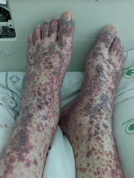
Eosinophilic granulomatosis with polyangiitis
Rare autoimmune vasculitis with three stages, formerly Churg–Strauss syndrome.

Eosinophilic granulomatosis with polyangiitis (EGPA) is an extremely rare autoimmune condition that causes inflammation of small and medium-sized blood vessels (vasculitis) in persons with a history of airway allergic hypersensitivity (atopy). It usually manifests in three stages: an early allergic stage, a stage of hypereosinophilia, and a vasculitic stage that can be life-threatening. The condition is now called EGPA to remove all eponyms from the vasculitides, having previously been known as Churg–Strauss syndrome.
Quick Facts
- Symptoms
- Fatigue, fever, weight loss, night sweats, abdominal pain, cough, joint pain, muscle pain, bleeding into tissues under the skin, a rash with hives, small bumps, or a general feeling of ill.
- Complications
- hypereosinophilia, granulomatosis, vasculitis, inner ear infections with fluid build up, inflammation of the moist membrane lining the surface of the eyelids, or inflammation of peripheral nerves.
- Risks
- History of allergy, asthma and asthma-associated lung abnormalities (i.e., pulmonary infiltrates).
- Treatment
- Suppress the activity of the immune system to alleviate inflammation.
- Medication
- Corticosteroid medications such as prednisone or methylprednisolone, and mepolizumab. Proliferation inhibitor for those with the presence of kidney or neurological disease.
Facts from the source article.
Lore & Background
Eosinophilic granulomatosis with polyangiitis (EGPA) was first described in 1951 by Jacob Churg and Lotte Strauss, who used the term allergic granulomatosis. It is a type of systemic necrotizing vasculitis. The condition is now called EGPA to remove all eponyms from the vasculitides; to facilitate the transition, it was referred to as 'eosinophilic granulomatosis with polyangiitis (Churg–Strauss)' for a period starting in 2012.
Reader's Guide
EGPA is a highly variable condition in terms of its presentation and course. The three stages—allergic, eosinophilic, and vasculitic—do not always occur in order, and some patients may develop severe or life-threatening complications such as gastrointestinal involvement and heart disease, while others are only mildly affected. The most serious complication of the vasculitic stage is heart disease, which causes nearly one-half of all deaths in EGPA patients. Diagnosis relies on criteria including asthma, eosinophilia, neuropathy, pulmonary infiltrates, sinus abnormalities, and histological evidence of extravascular eosinophils. Treatment involves glucocorticoids and other immunosuppressive drugs; in 2017, mepolizumab became the first drug specifically indicated for EGPA. The five-factor score stratifies risk of death based on renal function, proteinuria, gastrointestinal hemorrhage, central nervous system involvement, and cardiomyopathy.
Did You Know?
- Almost all patients experience asthma or allergic rhinitis, with more than 90% having a history of asthma.
- A normal 5% eosinophil composition in total leukocyte count can be elevated to 60% in EGPA.
- The ANCA+ subtype is characterized by predominantly vasculitis-like manifestations, while the ANCA− subtype is more commonly associated with eosinophilic-related symptoms.
- The FDA approved mepolizumab for EGPA on 12 December 2017.
The Patchwork of Symptoms and the Diagnostic Maze
GPA presents one of the most diagnostically challenging pictures in medicine because its opening signs are so nonspecific that patients often endure months or years of unexplained complaints before a unifying diagnosis emerges. In nearly every affected individual, the upper airway is the first site to show trouble—crusting around the nostrils, persistent congestion, recurrent nosebleeds, and a runny discharge that resists ordinary treatment. In some, erosion of the nasal septum produces the distinctive saddle-nose contour. Beyond the nose, the disease paints a remarkably broad canvas: more than half of patients develop scleritis, episcleritis, or conjunctivitis; roughly three-quarters who involve the kidneys suffer rapidly progressive glomerulonephritis that can march toward chronic renal failure; and the lungs may show coin-shaped nodules, infiltrates mistaken for pneumonia, cavitary lesions, or frank hemoptysis. Arthralgia or joint swelling appears in about sixty percent and is frequently misread as rheumatoid arthritis. Because no single symptom is pathognomonic, clinicians typically suspect GPA only after a prolonged trail of unexplained findings, and even tissue biopsy yields a definitive histological picture in only about half of cases.
The Immune Misfire: ANCAs, Neutrophils, and the Granuloma
The precise trigger that sets GPA in motion remains unidentified, yet the downstream immune choreography is increasingly well mapped. Antineutrophil cytoplasmic antibodies—specifically those directed against proteinase 3, an enzyme abundant in neutrophil granulocytes—are now widely regarded as the principal drivers of the vascular inflammation. Laboratory work has shown that these antibodies can switch neutrophils into an activated state, boost their stickiness to the inner lining of blood vessels, and provoke them to dump their granule contents, a cascade that tears through the endothelium and inflicts the most damage on small arterioles. Under the microscope, the classic triad of poorly formed granulomas, tissue necrosis, and multinucleated giant cells gives the disease its name, even though granulomas are not present in every biopsy. An intriguing hypothesis links chronic Staphylococcus aureus colonization of the nasal passages to the initial autoimmunity, suggesting that a persistent microbial stimulus may nudge the immune system toward self-attack. On the genetic side, variants in PTPN22, CTLA4, and human leukocyte antigen loci appear to modulate susceptibility, though the hereditary risk for any given individual stays low.
Taming the Storm: A Severity-Stratified Treatment Arsenal
Because GPA can range from a smoldering nasal irritation to a life-threatening multi-organ crisis, therapeutic strategy is calibrated to the intensity and organ involvement of each patient. In the most severe presentations, clinicians typically launch a two-pronged induction regimen: a potent immunosuppressant such as rituximab or cyclophosphamide paired with high-dose corticosteroids to rapidly quiet the inflammatory fire. Once the acute flare is controlled, the goal shifts to long-term remission maintenance, for which azathioprine, methotrexate, or a second course of rituximab are commonly employed to keep the disease in check without the heavier toxicity of induction agents. When the lungs, kidneys, or intestines sustain significant damage, plasma exchange is added to the protocol, physically removing circulating pathogenic antibodies and inflammatory mediators from the blood. The stakes of undertreatment are stark: involvement of the heart, lungs, or kidneys can prove fatal, and the rapidly progressive glomerulonephritis seen in three-quarters of kidney-affected patients can march inexorably toward chronic kidney disease if not arrested early. The overall approach thus balances aggressive immunosuppression against the risk of infection and other drug-related complications.
Who Gets It, Where It Lives, and How It Is Categorized
GPA is a genuinely rare condition. Across Europe, incidence estimates span from 2.1 to 14.4 new cases per million people per year, while in the United States the figure hovers around three per hundred thousand. The disease does not distribute evenly across populations: it is notably uncommon among Japanese and African-American communities but appears with greater frequency in individuals of Northern European ancestry. Men and women are affected at roughly equal rates, and pediatric cases are infrequently reported, making GPA predominantly an adult disorder. Taxonomically, GPA belongs to the family of ANCA-associated systemic vasculitides, a group that also includes eosinophilic granulomatosis with polyangiitis and microscopic polyangiitis. All three share the hallmark of circulating antineutrophil cytoplasmic antibodies attacking small- and medium-sized vessels, yet GPA is distinguished by its predilection for the ears, nose, and throat and by the presence of necrotizing granulomas. In the Chapel Hill classification system, despite its ability to involve medium-sized vessels, GPA is formally grouped among the small-vessel vasculitides. The condition was long known by the eponym Wegener's granulomatosis before the field adopted the current, more descriptive terminology.
Frequently Asked Questions
Who is Eosinophilic granulomatosis with polyangiitis?
EGPA is an ultra-rare autoimmune condition that attacks small and medium blood vessels, specifically in people who already carry a history of allergic airway hypersensitivity. Think of it as a three-act villain: it first stirs up allergy-like symptoms, then floods the bloodstream with eosinophils, and finally ignites destructive inflammation of the vessel walls.
What is Eosinophilic granulomatosis with polyangiitis known for?
Its signature move is a three-phase escalation—starting with an allergic phase, ramping into a hypereosinophilic phase, and culminating in a vasculitic phase where vessel walls become inflamed and can fail. This final stage is what makes the condition potentially fatal, as it can compromise multiple organ systems simultaneously.
Why is Eosinophilic granulomatosis with polyangiitis important?
EGPA sits at the crossroads of allergy, eosinophil biology, and systemic vasculitis, making it a key model for understanding how immune dysregulation can cascade from the airways all the way down to the vasculature. Its extreme rarity also means it is frequently misdiagnosed, so recognizing the three-stage pattern early is critical for timely treatment.
What is Eosinophilic granulomatosis with polyangiitis's backstory?
The condition was first described in 1951 by Jacob Churg and Lotte Strauss, who originally labeled it 'allergic granulomatosis.' It later became widely known as Churg–Strauss syndrome, and the eponym was eventually retired in favor of the descriptive, eponym-free label EGPA to align with the broader push to remove personal names from the vasculitis family.
More in Vasculitis 1-24
Spotted an error? Know more?
Reader corrections go straight into our review queue. Suggest an edit · How this site is sourced