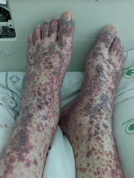
Cutaneous small-vessel vasculitis
Most common vasculitis; causes palpable purpura from small-vessel inflammation.

Cutaneous small-vessel vasculitis (CSVV) is inflammation of small blood vessels, usually accompanied by small lumps beneath the skin. It is the most common form of vasculitis seen in clinical practice, typically caused by inflammation of post-capillary venules in the dermis. The condition is also known by several other names, including hypersensitivity vasculitis and cutaneous leukocytoclastic vasculitis.
Quick Facts
- Field
- Rheumatology, Immunology
Facts from the source article.
Lore & Background
Cutaneous small-vessel vasculitis involves inflammation of small blood vessels in the skin, particularly post-capillary venules in the dermis. The term 'leukocytoclastic' refers to damage caused by nuclear debris from infiltrating neutrophils. Skin lesions initially appear as red to pink flat spots and raised bumps, then develop into non-blanching, palpable purpura—deep red to purple spots that feel raised and do not fade when pressed. Lesions are most commonly found symmetrically below the waist, especially on the buttocks and legs.
Reader's Guide
Cutaneous small-vessel vasculitis is significant as the most common vasculitis encountered in clinical practice. Its diagnosis is a diagnosis of exclusion, requiring ruling out systemic causes. A skin biopsy performed within 48 hours is the most definitive test. Treatment is directed at the underlying cause; if no cause is found and vasculitis is limited to the skin, supportive measures such as leg elevation and topical steroids are used. If it does not self-resolve within 3–4 weeks, oral colchicine or dapsone may be given. The condition can be persistent or recurrent, especially when associated with chronic connective tissue diseases.
Did You Know?
- 45–55% of cases are idiopathic, meaning the cause is unknown.
- Skin biopsy should be performed within 48 hours of appearance of the vasculitis for definitive diagnosis.
- Medications and infectious pathogens are the most common identifiable causes in adults, while IgA vasculitis frequently affects children.
Naming and Cultural Identity
This condition carries a remarkable collection of informal names that reflect its everyday origins rather than any formal medical taxonomy. It is most widely recognized as golfer's vasculitis, a label that stems from the extensive walking required during a round of golf—a pastime particularly popular among older adults. The moniker "Disney rash" emerged from the high frequency of cases among theme park visitors who spend long hours on their feet in warm weather. Other colloquial terms include hiker's rash, sport-induced vasculitis, and exercise-induced purpura, each pointing to a different recreational context in which the condition manifests. The naming pattern reveals that this is fundamentally a lifestyle-triggered phenomenon rather than a disease of a specific demographic. Cases have been linked not only to golf and hiking but also to cycling, dancing, swimming, and even prolonged outdoor cleaning. Despite its many colorful names, the underlying pathology remains the same: inflammation of small blood vessels in the lower legs. The condition is not contagious, and it disproportionately affects elderly individuals and those who are obese, making it a familiar but often underrecognized complaint in warm-weather recreational settings.
Development and Progression
The natural course of this condition follows a fairly predictable timeline from trigger to full recovery. The inciting event is typically a prolonged period of walking or standing in hot, humid conditions—golfing, sightseeing, or attending outdoor events are classic examples. Several hours after the activity, the first subtle sign appears: a feeling of warmth or mild discomfort in the lower legs. Within roughly 24 to 28 hours, the characteristic rash emerges as small reddish-purple spots or patches, which may gradually merge into larger areas of discoloration. Peak intensity generally arrives three to four days after onset, accompanied by the most noticeable itching, burning, and mild swelling. Without any specific medical intervention, symptoms begin to fade around the third or fourth day, with complete resolution typically occurring within seven to ten days, though some cases stretch to two or three weeks. A notable post-inflammatory feature is temporary hyperpigmentation, particularly in individuals with darker skin tones or those who have experienced repeated episodes; this darkening is not permanent and gradually fades. Recurrence is a hallmark of the condition—exposure to similar triggering circumstances can provoke another episode, and with repeated bouts, some individuals notice the rash developing more rapidly or with less intense provocation.
Clinical Presentation
The hallmark of this condition is a distinctive rash localized to the lower legs, typically appearing below the knees and above the ankles, though it can occasionally extend onto the top of the feet. The discoloration is described as reddish-purple, with a blotchy, mottled, or net-like reticular pattern that distinguishes it from other skin reactions. A key diagnostic clue is the distribution: the rash is usually bilateral and symmetrical, affecting both legs equally, and it characteristically spares areas covered by socks or shoes. Alongside the visible rash, patients commonly report mild to moderate itching, a burning or stinging sensation, and slight localized swelling. The affected skin may feel warm to the touch as a result of the underlying inflammatory process. Importantly, the condition remains strictly localized to the skin; it does not produce fever, joint pain, or other systemic symptoms that accompany many other forms of vasculitis. Mild pain or tenderness in the affected areas is less common but possible. In some individuals, especially those with darker complexions or a history of multiple episodes, the affected areas may develop temporary hyperpigmentation that persists for several weeks after the rash itself has cleared. The overall severity of these signs varies considerably from person to person.
Risk Factors and Mechanism
Although the exact pathophysiology behind this condition remains incompletely understood, preliminary research points to a convergence of environmental and physiological stressors. The primary risk factors involve unusual or prolonged physical activity—particularly weight-bearing, high-impact exercise—performed in hot and humid weather. The proposed mechanism involves two simultaneous processes: a spike in local tissue heat generated both by the external environment and by metabolic energy released in working muscles, and a pooling of blood in the lower extremities caused by prolonged upright posture. Together, these factors are thought to provoke localized inflammation of small capillaries in the legs. The condition is more frequently observed in elderly individuals and those who are obese, and it is not contagious. While golf and hiking are the most commonly cited triggers, documented cases have also followed cycling, dancing, swimming, and even extended outdoor cleaning tasks. A significant clinical consideration is the tendency toward recurrence: individuals who are repeatedly exposed to similar conditions—such as during hot seasons or vacations involving extensive walking—may experience multiple episodes. With each successive bout, some patients report that the rash appears more quickly or is provoked by less intense activity than before. The severity and duration of each episode can differ substantially between individuals, and not every person will progress through all stages in the same manner.
Frequently Asked Questions
Who is Cutaneous small-vessel vasculitis?
CSVV is the most frequently encountered vasculitis in everyday clinical practice, defined as an inflammatory assault on the tiny post-capillary venules within the dermis. It carries several aliases in the canon, most notably hypersensitivity vasculitis and cutaneous leukocytoclastic vasculitis.
What are Cutaneous small-vessel vasculitis's signature abilities?
Its hallmark presentation is non-blanching, palpable purpura—red-to-purple skin lesions that resist fading under pressure—often accompanied by itching, burning, joint aches, and a low-grade fever. It falls under the joint jurisdiction of Dermatology and Rheumatology.
How does Cutaneous small-vessel vasculitis's story typically resolve?
In most episodes the inflammation subsides once the offending trigger is removed or the immune response settles, but confirming the diagnosis reliably requires a skin biopsy performed within the first 48 hours. It is ultimately a diagnosis of exclusion, so clinicians must rule out other mimics before locking in the label.
Why is Cutaneous small-vessel vasculitis important?
Because it is the single most common vasculitis seen in practice, it acts as the primary entry point for recognizing small-vessel inflammation and can serve as the first visible clue to an underlying autoimmune condition, infection, or drug reaction. Its sheer prevalence makes it the reference standard against which rarer vasculitides are compared.
What triggered Cutaneous small-vessel vasculitis's appearance?
Known inciting factors include certain medications, infections, allergens, and autoimmune conditions, yet in roughly 45 to 55 percent of cases no identifiable cause is ever found, leaving the episode classified as idiopathic. This unresolved origin is a recurring and frustrating thread in its clinical narrative.
More in Vasculitis 1-24
Spotted an error? Know more?
Reader corrections go straight into our review queue. Suggest an edit · How this site is sourced