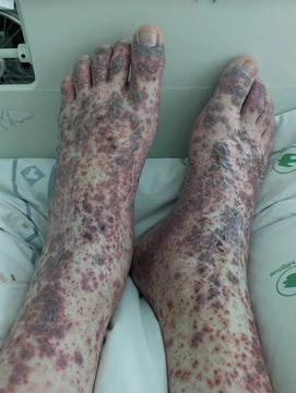
Arteritis
Vascular disorder involving inflammation of artery walls.

Arteritis is a condition where the walls of arteries become inflamed, often due to an infection or an autoimmune reaction. This complex disorder is not yet fully understood. It can be categorized into different types depending on which organ systems are involved. A serious potential complication is thrombosis, which can be fatal. Arteritis, along with phlebitis, falls under the broader category of vasculitis. General signs of arteritis can include inflammation, fever, anemia of chronic disease (rather than increased red blood cell production), limping, and a reduced pulse. Doctors diagnose arteritis based on unusual medical symptoms. These symptoms can resemble those of other conditions, such as Ehlers–Danlos syndrome, Marfan syndrome, tuberculosis, syphilis, spondyloarthropathies, Buerger's disease, Behçet's disease, and Kawasaki disease. Several imaging techniques are used to diagnose and track the disease. These include direct angiography, magnetic resonance angiography (MRA), and ultrasonography. Angiography is often used for Takayasu arteritis, especially in advanced stages where arterial narrowing, blockages, and aneurysms can be seen. However, it is invasive and exposes patients to high radiation, so it is not recommended for routine long-term monitoring. Computed tomography angiography (CTA) can measure the aorta and its branches and identify vessel wall lesions in middle to late stages, also showing blood flow, but it too involves high radiation exposure. MRA is useful for diagnosing Takayasu arteritis in early stages, showing changes like vessel wall thickening. It can detect even small changes and is good for monitoring without radiation, but it is expensive and shows calcification less clearly than other methods. Ultrasonography is ideal for early diagnosis when inflammation is present, and it can show blood flow. It is a popular first-line test because it is quick, cheap, noninvasive, and radiation-free, and it is used for long-term monitoring. However, not all vascular lesions are visible on ultrasound, and accuracy depends on the person performing the scan. Arteritis can be primary or secondary to another disease. An example of secondary arteritis is that caused by certain infections, though *Candida albicans* is not a standard or widely recognized cause in medical literature.
- Field
- Vascular medicine
- Known for
- Inflammation of artery walls, including types such as giant cell arteritis, Takayasu arteritis, and temporal arteritis
- Types
- Primary and secondary arteritis
- Complication
- Thrombosis (can be fatal)
Lore & Background
Arteritis is a vascular disorder characterized by inflammation of the walls of arteries, usually as a result of infection or autoimmune responses. It is a complex disorder still not entirely understood. Arteritis may be distinguished by its different types, based on the organ systems affected. A complication of arteritis is thrombosis, which can be fatal. Arteritis and phlebitis are forms of vasculitis.
Signs of general arteritis may include inflammation, fever, anemia of chronic disease (not increased red blood cell production), limping, and reduced pulse. Diagnosis is based on unusual medical symptoms, and similar symptoms may be caused by other conditions such as Ehlers–Danlos syndrome, Marfan syndrome, tuberculosis, syphilis, spondyloarthropathies, Buerger's disease, Behçet's disease, and Kawasaki disease.
Reader's Guide
Arteritis is significant as a vascular disorder that can lead to serious complications such as thrombosis. Its classification into primary and secondary types, and further into specific forms like giant cell arteritis, Takayasu arteritis, and temporal arteritis, helps guide diagnosis and treatment. The condition is diagnosed through imaging techniques, each with advantages and limitations: angiography is invasive and uses high radiation, while ultrasonography is noninvasive and suitable for early stages and long-term monitoring. Treatment primarily involves glucocorticoids, with cytotoxic medications as adjuncts. The legacy of arteritis research lies in understanding its inflammatory mechanisms and improving diagnostic and therapeutic approaches, though much remains unknown about its underlying causes.
Did You Know?
- Takayasu arteritis is most common in females, with a median age of 25 years.
- Temporal arteritis has a higher incidence in people of Scandinavian descent.
Defining Arteritis Within the Vasculitis Family
Arteritis sits within the broader family of vasculitis—disorders in which inflammation systematically destroys the walls of blood vessels. While the term vasculitis encompasses damage to both arteries and veins, arteritis specifically refers to the inflammatory targeting of arterial tissue, making it a distinct clinical entity from phlebitis, the venous counterpart. The underlying mechanism in most cases centers on leukocyte migration: white blood cells infiltrate the vessel wall, and the resulting immune-mediated damage progressively compromises vascular integrity. Lymphangitis, the inflammation of lymphatic vessels, is occasionally grouped under the vasculitis umbrella, though it involves a different vascular network entirely. What makes arteritis particularly challenging is that it does not present as a single disease but rather as a spectrum of conditions, each shaped by which vessels are targeted, how severely, and what triggered the inflammatory cascade. Understanding arteritis therefore requires distinguishing it from its venous counterpart while recognizing that the two can coexist within the same patient, compounding both the diagnostic and therapeutic picture.
Clinical Presentation: What Patients Experience
The outward signs of arteritis and related vasculitides are heavily influenced by the caliber of the vessels primarily involved, meaning two patients with technically the same diagnosis can look remarkably different depending on whether large, medium, or small arteries bear the brunt of the inflammation. That said, a constellation of non-specific systemic symptoms appears across virtually every form: persistent fever, headaches, profound fatigue, muscle aches, unexplained weight loss, and joint pain. These vague complaints often delay recognition because they mimic far more common ailments. On the skin, however, the picture becomes more distinctive. Even large-vessel vasculitides can produce cutaneous findings, and the most frequently observed manifestations include purpura, firm nodules, a mottled pattern known as livedo reticularis, open skin ulcers, and purpuric urticaria. The diversity of these presentations means clinicians must correlate vascular size involvement with the specific organ and tissue damage they observe, rather than relying on any single hallmark sign.
The Chapel Hill Classification: Mapping the Spectrum
The 2012 Chapel Hill Consensus Conference established the nomenclature framework that organizes vasculitis into primary systemic, secondary, and single-organ categories, with primary systemic further subdivided by vessel caliber. Large-vessel vasculitis, which preferentially targets the aorta and its major branches, includes Takayasu arteritis and giant cell arteritis. Medium-vessel disease, affecting the arteries that supply internal organs, encompasses polyarteritis nodosa and Kawasaki disease. Small-vessel vasculitis splits into two major branches: ANCA-associated forms (eosinophilic granulomatosis with polyangiitis, granulomatosis with polyangiitis, and microscopic polyangiitis) and immune-complex forms (including cryoglobulinemic vasculitis, IgA vasculitis, anti-C1q vasculitis, and anti-GBM disease). Variable-vessel vasculitis, which can strike arteries, veins, and capillaries of any size without a predominant target, includes Behçet's disease and Cogan's syndrome. Secondary and single-organ forms round out the taxonomy, the latter confined to one organ system such as the gut, skin, or peripheral nerves.
Diagnosis: From Bloodwork to Biopsy
Confirming arteritis or any vasculitis demands a layered diagnostic approach. Initial laboratory work on blood or body fluids typically reveals markers of active systemic inflammation: a raised erythrocyte sedimentation rate, elevated C-reactive protein, anemia, a heightened white blood cell count, and sometimes eosinophilia. Depending on the subtype, antineutrophil cytoplasmic antibody levels may be elevated, and hematuria can point to renal involvement. Organ-specific functional tests may also show abnormalities proportional to the degree of tissue damage, and imaging such as a brain SPECT can document reduced cerebral perfusion. The gold standard, however, remains tissue biopsy of the affected organ—skin, sinus, lung, nerve, brain, or kidney—because it reveals the precise pattern of vascular inflammation. In certain subtypes, pathologists observe leukocytoclasis, where nuclear debris from infiltrating neutrophils fragments the vessel wall, often manifesting clinically as palpable purpura. When biopsy is impractical, angiography or 18F-fluorodeoxyglucose PET/CT scanning can demonstrate characteristic inflammatory patterns in the vasculature.
Frequently Asked Questions
Who is Arteritis?
Arteritis is a vascular condition in which arterial walls develop inflammation, usually triggered by an infectious agent or the body's own immune system misdirected against its own vessels. It sits within the broader family of vasculitis disorders.
What is Arteritis known for?
Its primary 'ability' is to cause swelling and thickening of arterial walls, narrowing the passage for blood flow. This can produce symptoms such as persistent fever, unexplained weight loss, a shuffling gait, and diminished pulse in affected limbs.
Why is Arteritis important?
It is clinically significant because it can affect virtually any organ system and presents in multiple distinct forms, such as giant cell arteritis, Takayasu arteritis, and temporal arteritis. Early recognition is critical because the risk of catastrophic clotting rises the longer the inflammation goes untreated.
How does Arteritis relate to Phlebitis?
Both are siblings under the umbrella term 'vasculitis,' but Arteritis targets arterial walls while Phlebitis focuses on veins. Understanding which vessel type is involved helps clinicians narrow the diagnosis and choose the right therapeutic path.
More in Vasculitis 1-24
Spotted an error? Know more?
Reader corrections go straight into our review queue. Suggest an edit · How this site is sourced