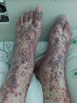
Anti-neutrophil cytoplasmic antibody
Autoantibodies linked to small-vessel vasculitis.

Anti-neutrophil cytoplasmic antibodies (ANCAs) are a group of autoantibodies, mainly of the IgG type, directed against antigens in the cytoplasm of neutrophils and monocytes. They are detected through blood tests and are particularly associated with systemic vasculitides known as ANCA-associated vasculitides (AAV).
- Field
- Immunology, Autoimmune Disease
- Known for
- Association with systemic vasculitis (ANCA-associated vasculitides)
- Detection methods
- Immunofluorescence (IF) and Enzyme-linked immunosorbent assay (ELISA)
- Main antigens
- Proteinase 3 (PR3) and Myeloperoxidase (MPO)
Lore & Background
ANCAs are detected using immunofluorescence on ethanol-fixed neutrophils, revealing four patterns: cytoplasmic (c-ANCA), atypical C-ANCA, perinuclear (p-ANCA), and atypical ANCA (a-ANCA). The c-ANCA antigen is specifically proteinase 3 (PR3), while p-ANCA antigens include myeloperoxidase (MPO) and bactericidal/permeability-increasing protein (BPI). ELISA is used to confirm antibodies to individual antigens, most commonly MPO and PR3.
The development of ANCAs is poorly understood, with hypotheses including molecular mimicry—where microbial superantigens resemble self-antigens—and defective apoptosis, where ineffective removal of apoptotic neutrophil fragments exposes intracellular molecules to the immune system. Genetic susceptibility and environmental factors, such as exposure to silicates, are thought to contribute.
ANCAs are associated with small-vessel vasculitides including granulomatosis with polyangiitis, microscopic polyangiitis, and eosinophilic granulomatosis with polyangiitis. PR3-directed c-ANCA is present in 80-90% of granulomatosis with polyangiitis, while MPO-specific p-ANCA is found in 50% of microscopic polyangiitis. The presence or absence of ANCA cannot alone indicate disease, and their role in pathogenesis remains controversial, though in vitro and animal models support a direct pathological role via neutrophil activation and endothelial damage.
Reader's Guide
Anti-neutrophil cytoplasmic antibodies are significant as serological markers for a group of life-threatening systemic vasculitides, enabling diagnosis and classification of conditions such as granulomatosis with polyangiitis and microscopic polyangiitis. Their detection via immunofluorescence and ELISA is standard in clinical laboratories. Despite ongoing debate about their exact pathogenic role, evidence from animal models and in vitro studies suggests ANCAs can directly activate neutrophils, leading to endothelial injury through degranulation and release of lytic enzymes. The reappearance of ANCAs after treatment may indicate relapse, though titers do not always correlate with disease activity. The discovery that up to 70% of patients with granulomatosis with polyangiitis are chronic nasal carriers of Staphylococcus aureus supports the molecular mimicry hypothesis. Treatment with rituximab, which depletes B-cells, can induce remission even without a decrease in ANCA titers, suggesting a direct role for ANCA-reactive B-cells. Overall, ANCAs remain a critical focus of research in autoimmune vasculitis.
Did You Know?
- c-ANCA is specifically directed against proteinase 3 (PR3) and is present in 80-90% of granulomatosis with polyangiitis.
- p-ANCA without nuclear extension occurs with antibodies to BPI, cathepsin G, elastase, lactoferrin, and lysozyme.
- Levamisole, a common adulterant of cocaine, can cause an ANCA-positive vasculitis.
Visual Classification Through Immunofluorescence
ANCAs are first identified in the laboratory through indirect immunofluorescence performed on ethanol-fixed neutrophils, though formalin-fixed cells can be substituted to sharpen the distinction between pattern types. Under the microscope, the staining topology falls into four broad categories. The classic cytoplasmic pattern, abbreviated c-ANCA, produces a granular glow throughout the cell body with a characteristic central accentuation along interlobular regions. An atypical variant, sometimes written C-ANCA, yields a more uniform cytoplasmic stain lacking that interlobular emphasis. The perinuclear pattern, p-ANCA, splits into three subtypes: classical p-ANCA where fluorescence hugs the nucleus and extends into it, a variant without nuclear extension that stays strictly perinuclear, and a granulocyte-specific antinuclear antibody form that lights up only the nuclei of granulocytes. Finally, the atypical or x-ANCA pattern is the most heterogeneous, frequently displaying both cytoplasmic and perinuclear fluorescence simultaneously. These visual distinctions guide clinicians toward the correct antigenic target before confirmatory testing.
Antigenic Targets and Confirmatory Assays
Once a fluorescent pattern is observed, the laboratory moves to enzyme-linked immunosorbent assay to pin down the exact protein being attacked. The two most frequently tested targets on a microtitre plate are proteinase 3, the hallmark antigen of classic c-ANCA, and myeloperoxidase, the principal target of classical p-ANCA. Beyond these two, the antigenic landscape is considerably wider. p-ANCA without nuclear extension can be directed against bactericidal/permeability-increasing protein, cathepsin G, elastase, lactoferrin, or lysozyme. Granulocyte-specific antinuclear antibodies recognize nuclear antigens unique to granulocytes. Atypical ANCA antigens overlap with p-ANCA targets but may reflect differences in how neutrophils are processed during fixation. A long tail of less common targets has also been catalogued, including HMG1, HMG2, alpha enolase, catalase, beta glucuronidase, azurocidin, actin, and h-lamp-2, each producing either a perinuclear, cytoplasmic, or mixed staining profile. In cystic fibrosis, for instance, atypical c-ANCA predominantly targets BPI, illustrating how the same antibody class can point to very different underlying conditions depending on which intracellular protein it recognizes.
How These Autoantibodies Arise
The precise origin of ANCA remains one of the open questions in autoimmunity. Researchers agree that a genetic predisposition, likely involving genes that modulate the intensity of immune responses, interacts with environmental triggers such as silicate exposure, yet no single pathway fully explains how the immune system begins targeting intracellular proteins. Two leading hypotheses have emerged. The molecular mimicry theory proposes that microbial superantigens, particularly those from Staphylococcus and Streptococcus species, carry structural regions resembling self-antigens, thereby cross-activating T-cells and igniting a residual autoimmune attack. This is supported by the observation that up to seventy percent of patients with granulomatosis with polyangiitis are chronic nasal carriers of Staphylococcus aureus, and those carriers face roughly an eight-fold increased risk of disease relapse, consistent with a type II hypersensitivity mechanism. The defective apoptosis theory offers a complementary explanation: when neutrophils fail to undergo programmed cell death properly, or when apoptotic debris is incompletely cleared, intracellular molecules normally hidden from the immune system become exposed, providing the antigenic stimulus that drives ANCA production. Neither theory yet accounts for why different patients develop different antigen specificities, and research into ANCA pathogenesis continues.
Clinical Landscape and Disease Associations
ANCAs are most tightly linked to the family of small-vessel vasculitides collectively termed ANCA-associated vasculitides, a group that received updated classification criteria in 2022. Proteinase 3–directed c-ANCA appears in roughly eighty to ninety percent of granulomatosis with polyangiitis cases, in twenty to forty percent of microscopic polyangiitis, in a similar proportion of primary pauci-immune necrotizing crescentic glomerulonephritis, and in about a third of eosinophilic granulomatosis with polyangiitis. Myeloperoxidase-specific p-ANCA, by contrast, is detected in approximately half of microscopic polyangiitis and pauci-immune glomerulonephritis patients and in roughly a third of eosinophilic granulomatosis with polyangiitis. Atypical c-ANCA targeting BPI is found in about eighty percent of cystic fibrosis patients and also in inflammatory bowel disease, primary sclerosing cholangitis, and rheumatoid arthritis. Drug-induced vasculitis, autoimmune liver disease, parasitic infections, and even levamisole-contaminated cocaine can all generate ANCA-positive results. Importantly, the mere presence or absence of these antibodies cannot confirm or exclude a diagnosis; results must always be interpreted alongside the full clinical picture, and the correlation between ANCA levels and disease activity remains a subject of ongoing debate.
Frequently Asked Questions
Who is Anti-neutrophil cytoplasmic antibody?
ANCA is not a single character but a family of autoantibodies, mostly IgG in class, that mistakenly lock onto proteins housed inside the cytoplasm of neutrophils and monocytes. They belong to the immunology and autoimmune-disease canon and are best known for their link to small-vessel inflammation.
What is Anti-neutrophil cytoplasmic antibody known for?
The two principal targets in ANCA's 'arsenal' are Proteinase 3 (PR3) and Myeloperoxidase (MPO), both granule-resident enzymes in neutrophils. By activating these cells and amplifying inflammatory signaling, ANCA drives the tissue-damaging pattern seen in ANCA-associated vasculitides (AAV).
How do you detect Anti-neutrophil cytoplasmic antibody?
Two standard blood-test methods are used: indirect immunofluorescence (IF), which reveals a characteristic staining pattern on ethanol-fixed neutrophils, and ELISA, which quantifies antibodies against specific antigens such as PR3 or MPO. Together they confirm both the presence and the subtype of the antibody.
Why is Anti-neutrophil cytoplasmic antibody important?
ANCA is the key diagnostic flag that separates the ANCA-associated vasculitides from other small-vessel vasculitides, directly shaping how clinicians stage disease and choose immunosuppressive therapy. Without this serological marker, distinguishing conditions like granulomatosis with polyangiitis from microscopic polyangiitis would be far more difficult.
More in Vasculitis 1-24
Spotted an error? Know more?
Reader corrections go straight into our review queue. Suggest an edit · How this site is sourced