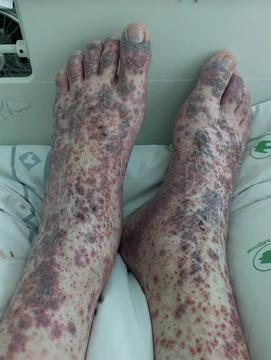
Microscopic polyangiitis
Autoimmune small-vessel vasculitis without granulomatous inflammation.

Microscopic polyangiitis is an autoimmune condition in which the body attacks its own small blood vessels, causing inflammation and tissue death. Unlike some similar diseases, it does not involve the formation of granulomas. It belongs to a group of disorders known as ANCA-associated vasculitides.
The illness can produce general symptoms such as fever, joint and muscle pain, loss of appetite, weight loss, and fatigue. Because it can affect many organs, the specific signs vary widely. Patients may experience cough, shortness of breath, or coughing up blood. Kidney problems occur in up to 80% of cases, showing up as blood and protein in the urine, which can lead to either rapid or gradual kidney failure. Lung involvement, seen in 20–50% of patients, can cause bleeding in the lungs or chronic scarring that leads to respiratory failure. Skin changes like palpable purpura or livedo racemosa, seizures or peripheral nerve damage, and abdominal pain are also possible.
The exact cause is not fully understood. The leading idea is that people with a genetic predisposition develop the disease when an unknown trigger prompts the production of p-ANCA antibodies. These antibodies circulate at low levels until an environmental trigger—such as an infection, cancer, or certain drugs—causes an increase in neutrophils. The neutrophils bind to the p-ANCAs and release inflammatory chemicals, reactive oxygen molecules, and enzymes that damage the inner lining of small blood vessels, leading to inflammation and necrosis. In the kidneys, this damage is specifically called necrotizing and crescentic glomerulonephritis.
Diagnosis often starts with lab tests showing a high sedimentation rate, elevated C-reactive protein, and anemia. Kidney impairment raises blood creatinine levels and brings protein and red blood cells into the urine. A key test detects perinuclear antineutrophil cytoplasmic antibodies (p-ANCA) that target myeloperoxidase. Depending on the organs involved, additional tests may include a kidney biopsy or electromyography for nerve damage.
The symptoms can resemble those of granulomatosis with polyangiitis (GPA), another small-vessel vasculitis. However, microscopic polyangiitis typically lacks the significant upper respiratory tract problems—like sinusitis—that are common in GPA.
Standard treatment aims to stop blood vessel inflammation and achieve remission.
- Field
- Autoimmune disease / Vasculitis
- Known for
- Systemic, pauci-immune, necrotizing small-vessel vasculitis without granulomatous inflammation
- Affected organs
- Kidneys (up to 80% of cases), lungs (20-50% of cases), skin, nervous system
Lore & Background
Microscopic polyangiitis presents with a wide range of clinical features, including constitutional symptoms such as fever, arthralgia, myalgia, loss of appetite, weight loss, and fatigue. Organ-specific manifestations include cough, shortness of breath, hemoptysis, kidney failure, skin manifestations like palpable purpura and livedo racemosa, seizures, peripheral neuropathy, and abdominal pain. The kidneys are affected in up to 80% of cases, with signs of blood and protein in the urine, potentially leading to rapidly or slowly progressive kidney failure. The lungs are affected in 20-50% of cases, with findings of pulmonary hemorrhage or chronic pulmonary fibrosis leading to respiratory failure.
Reader's Guide
The leading hypothesis for the cause of microscopic polyangiitis is that ANCA-associated vasculitis develops in patients with a genetic predisposition when an unknown trigger causes production of p-ANCA. These antibodies circulate at low levels until an environmental trigger—such as infection, malignancy, or drug therapy—causes upregulation of neutrophils. The neutrophils bind to p-ANCAs and release inflammatory cytokines, reactive oxygen species, and lytic enzymes that cause endothelial injury, resulting in inflammation and necrosis of small vessels. Kidney damage is specifically called necrotizing and crescentic glomerulonephritis. Diagnosis relies on laboratory tests showing elevated sedimentation rate, CRP, anemia, elevated creatinine, and protein and red blood cells in urine, along with detection of p-ANCA with myeloperoxidase specificity. Special tests such as renal biopsy or electromyography may be performed. The differential diagnosis includes granulomatosis with polyangiitis, but microscopic polyangiitis typically lacks significant upper respiratory tract involvement. Treatment involves immunosuppressive therapy with high-dose glucocorticoids combined with either cyclophosphamide or rituximab; plasmapheresis may be used in life-threatening cases. Therapy is tapered over months, but discontinuation can increase risk of disease flares.
Did You Know?
- Microscopic polyangiitis is characterized by a systemic, pauci-immune, necrotizing, small-vessel vasculitis without granulomatous inflammation.
- The leading hypothesis involves p-ANCA antibodies that bind to neutrophils, causing endothelial injury.
More in Vasculitis 1-24
Spotted an error? Know more?
Reader corrections go straight into our review queue. Suggest an edit · How this site is sourced