Sialadenitis
Inflammation of salivary glands, often from infection, stones, or autoimmune disease.
Sialadenitis is a condition where the salivary glands become inflamed, most often the parotid, then the submandibular and sublingual. It differs from sialadenosis, a non-inflammatory gland enlargement. Cases are either acute or chronic, and triggers include bacterial, viral, and autoimmune causes.
Quick Facts
- Most common gland affected
- Parotid gland
Facts from the source article.
Did You Know?
- Recurrent parotitis of childhood is characterized by periods of pain and swelling in the parotid gland accompanied by fever.
- Chronic sclerosing sialadenitis is commonly unilateral and can mimic a tumour.
- In chronic recurrent sialadenitis, acute attacks are managed with hydration, NSAIDs, sialogogues, and gentle gland massage; surgical excision is considered if attacks occur more than about three times per year.
Causes
Sialadenitis can arise from cancer, autoimmune conditions, viral and bacterial infections, idiopathic causes, or stones formed mainly from calculus. Morphological characteristics of salivary ducts were once thought to contribute, as saliva stagnation might increase incidence, but a small study of 106 found no statistically significant difference in duct length or angles and the likelihood of developing sialadenitis; it also confirmed that age, gender, side of face, and degree of sialadenitis had no impact on these duct features. Viral pathogens cause sialadenitis more often than bacterial ones. Mumps is the most common virus affecting the parotid and submandibular glands, with the parotid most frequently involved. Other viruses include HIV, coxsackie, and parainfluenza; HIV parotitis is classically asymptomatic or a non-painful swelling. Common bacterial causes are Staphylococcus aureus, Streptococcus pyogenes, viridans streptococci, and Haemophilus influenzae. Autoimmune causes include Sjögren's syndrome, sarcoidosis, and granulomatosis with polyangiitis. Sjögren's syndrome and sarcoidosis are the most common causes of chronic sialadenitis and are often considered primary, though other contributing factors may be present. Heerfordt's syndrome, a form of sarcoidosis, features facial nerve palsy, parotid enlargement, and anterior uveitis. One study found that salivary calculi were the main indicator for submandibular gland removal when neoplasia was absent, as 82% of removed glands contained calculi and all but one of those had chronic sialadenitis.
Histopathology
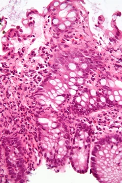
In acute bacterial sialadenitis, the initial stage involves accumulation of bacteria, neutrophils, and inspissated fluid in ductal lumens. Damage to ductal epithelium leads to sialodochitis (periductal inflammation), neutrophil accumulation in glandular stroma, and acini necrosis with microabscess formation. Recurrent episodes produce chronic sialadenitis, with periductal lymph follicles and further acinar destruction. In acute bacterial and viral cases, lobular architecture is maintained or slightly expanded; areas of liquefaction may indicate abscess. Acute bacterial sialadenitis shows acinar destruction with interstitial neutrophil infiltrates and small abscesses with necrosis. Viral sialadenitis displays vacuolar changes in acini with lymphocytic and monocytic interstitial infiltrate; cytomegalovirus sialadenitis may be asymptomatic. Chronic sialadenitis (lymphoepithelial sialadenitis) has 50% monoclonal by PCR, while MALT lymphoma shows ducts surrounded by broad coronas of monocytoid cells, infiltration of interfollicular regions, atypical plasma cells with Dutcher bodies, monoclonality by immunohistochemistry or flow cytometry, and monocytoid infiltrates in regional lymph nodes. Histologically, chronic sialadenitis ranges from unremarkable to firm tan with lobular expansion or atrophy. Salivary stones (sialoliths) may be present with cystic duct dilation and periductal fibrosis; mucus extravasation may occur. Common observations include chronic inflammatory infiltrate (lymphocytes, plasma cells, macrophages), fibrosis, acinar atrophy, and mucous cell metaplasia of ducts. In sialolithiasis, squamous metaplasia and dark calcific stone fragments may be seen.
Diagnosis
Diagnosis of sialadenitis involves considering history, presenting signs and symptoms, and appropriate investigations, along with the type of gland affected and underlying conditions such as autoimmune disease or ductal stones. There may be a history of recent surgery or use of medications like antihistamines, antidepressants, or anticholinergic agents, which can cause objective hypofunction or subjective dry mouth. A decrease in salivary volume from systemic disease may also be reported. Physical examination may reveal unilateral or bilateral painful swelling of the parotid or submandibular regions, sometimes with external displacement of the earlobe adjacent to an inflamed parotid. Pus may discharge from major salivary duct openings spontaneously or after gland manipulation. Mandibular trismus is rare but possible with larger swellings; dysphagia may occur. Fever may be present, and spiking temperatures suggest abscess formation. Chronic recurrent episodes may indicate underlying Sjögren's syndrome or ductal abnormalities, with a prodrome of tingling before pain and swelling. Chronic sclerosing sialadenitis typically presents as a unilateral submandibular swelling indistinguishable from a neoplasm, with pain inconsistent. Sialoliths cause obstructive sialadenitis, with abrupt episodic swelling around meal times lasting 2–3 hours. Persistent, asymptomatic bilateral parotid swelling may represent systemic disease; dry eyes and dry mouth suggest connective tissue disease such as systemic lupus erythematosus, rheumatoid arthritis, or scleroderma. Oral candidiasis may also be present.
Epidemiology
Sialadenitis of the parotid gland accounts for a much larger proportion of hospital admissions than submandibular sialadenitis, which represents only 10% of diagnosed cases. Chronic sialadenitis is relatively common, while bacterial sialadenitis and sclerosing polycystic sialadenitis are rare. Chronic sclerosing sialadenitis predominantly affects males over 50, with 40% of cases having an allergic disease such as chronic sinusitis or bronchial asthma. One study of 112 patients from England and Wales, aged 12 to 81, reported a mean age of 39 years (standard deviation 16). Most patients underwent surgery between ages 20 and 69, but symptoms most often began between ages 20 and 49. In each age group, most patients were in their twenties; a significant number of females had symptom onset in their thirties and forties. More women than men reported symptoms confirmed as sialadenitis, suggesting a possible female predominance, though further research is needed. In the United States, acute suppurative parotitis accounts for 0.01–0.02% of hospital admissions, with the submandibular gland involved in 10% of major salivary gland cases. No predilection for race, sex, or age was found, though sialadenitis tended to occur in debilitated, dehydrated, or older individuals. Bacterial sialadenitis is now uncommon and usually associated with sialoliths. In the United Kingdom, the incidence of admissions for sialadenitis is 27.5 per million population, with mumps the most common cause. Ascending acute bacterial parotitis, once a common perimortal event, is now rare due to antibiotics and modern care reducing dehydration.
More in Inflammatory Diseases of the Digestive System
Spotted an error? Know more?
Reader corrections go straight into our review queue. Suggest an edit · How this site is sourced