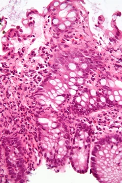
Ascending cholangitis
Inflammation of the bile duct, often due to gallstone obstruction.
Ascending cholangitis, or acute cholangitis, is a bile duct infection. Bacteria from the small intestine travel upward into the duct, often when gallstones have already caused a partial blockage. This is a life-threatening medical emergency. Symptoms include fever, abdominal pain, and yellowing of the skin or eyes. Treatment starts with intravenous fluids and antibiotics, and an endoscopy may be needed to clear the obstruction.
Quick Facts
- Field
- Gastroenterology; General surgery
- Symptoms
- jaundice, fever and abdominal pain
Facts from the source article.
Did You Know?
- Reynolds' pentad includes Charcot's triad plus septic shock and mental confusion, indicating worsening and sepsis.
- Biliary sepsis is a systemic complication of acute cholangitis when infection spreads from the bile ducts into the bloodstream.
Causes
Bile duct obstruction in acute cholangitis is generally due to gallstones, but 10–30% of cases arise from other causes such as benign stricturing, postoperative damage, altered bile duct structure, or various tumors including cancer of the bile duct, gallbladder, ampulla of Vater, pancreas, or duodenum. Anaerobic organisms like Clostridium and Bacteroides may be involved, especially in the elderly and those with prior biliary surgery. Parasites such as Ascaris lumbricoides and liver flukes (Clonorchis sinensis, Opisthorchis viverrini, Opisthorchis felineus) can cause cholangitis. In people with AIDS, opportunistic organisms have caused AIDS cholangiopathy, though risk has diminished with effective treatment. Cholangitis may also complicate ERCP; prophylactic antibiotics are recommended for those undergoing this procedure. A permanent biliary stent slightly increases risk.
Pathogenesis
Bile is produced by the liver, stored and concentrated in the gallbladder, and released into the duodenum through the common bile duct and ampulla of Vater, controlled by the sphincter of Oddi. The biliary tree is normally relatively free of bacteria due to protective mechanisms: the sphincter of Oddi acts as a mechanical barrier, low pressure (8 to 12 cmH2O) allows free bile flow, and continuous forward flow flushes bacteria into the duodenum. Bile salts and immunoglobulin secreted by the bile duct epithelium also have a protective role. Bacterial contamination alone without obstruction usually does not cause cholangitis. However, increased pressure within the biliary system (above 20 cmH2O) from obstruction widens spaces between lining cells, bringing bacterially contaminated bile into contact with the bloodstream. It also adversely affects Kupffer cells, which are specialized macrophages that help prevent bacteria from entering the biliary system, and decreases production of IgA immunoglobulins in bile. This results in bacteremia and the systemic inflammatory response syndrome (SIRS), which in the presence of suspected or confirmed infection is called sepsis. Biliary obstruction itself impairs immune function by affecting neutrophil granulocytes and modifying cytokine levels.
Diagnosis
Routine blood tests show features of acute inflammation, such as raised white blood cell count and elevated C-reactive protein, and usually abnormal liver function tests. In most cases, liver function tests indicate obstruction with raised bilirubin, alkaline phosphatase, and γ-glutamyl transpeptidase; early on, however, they may resemble hepatitis with elevations in alanine transaminase and aspartate transaminase. Blood cultures yield the causative bacteria in 36% of cases, usually after 24–48 hours of incubation. The most common bacteria are gram-negative bacilli: Escherichia coli (25–50%), Klebsiella (15–20%), and Enterobacter (5–10%); among gram-positive cocci, Enterococcus causes 10–20%. Medical imaging is used to identify the site and nature of obstruction. Ultrasound is often the first investigation, showing bile duct dilation and identifying 38% of bile duct stones, though it is poor at identifying stones farther down the duct. Magnetic resonance cholangiopancreatography (MRCP) has comparable sensitivity to ERCP, but smaller stones may still be missed depending on facility quality.
Treatment
Cholangitis requires hospital admission. Intravenous fluids are administered, especially if blood pressure is low, and antibiotics are commenced. Empirical broad-spectrum antibiotics are used until the pathogen and its sensitivities are known. Combinations of penicillins and aminoglycosides are widely used; ciprofloxacin is effective in most cases and may be preferred due to fewer side effects. Metronidazole is often added for anaerobic pathogens, particularly in very ill or at-risk patients. Antibiotics are continued for 7–10 days. Vasopressors may be required to counter low blood pressure. Definitive treatment is relief of the underlying biliary obstruction, usually deferred 24–48 hours until the patient is stable, but may be done emergently if deterioration continues despite treatment or if antibiotics are ineffective (in 15% of cases). Endoscopic retrograde cholangiopancreatography (ERCP) is the most common approach, involving endoscopy to identify the ampulla of Vater and insert a tube into the bile duct. Sphincterotomy is typically performed to ease bile flow and allow instrument insertion for stone extraction; the duct orifice may also be dilated with a balloon. Stones are removed by suction or instruments such as balloons and baskets. Larger stones may require a mechanical lithotriptor or extracorporeal shock wave lithotripsy.
More in Inflammatory Diseases of the Digestive System
Spotted an error? Know more?
Reader corrections go straight into our review queue. Suggest an edit · How this site is sourced