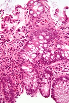
Acute pancreatitis
Sudden pancreatic inflammation with high mortality in severe cases.
Acute pancreatitis is a sudden inflammation of the pancreas that can occur as a single episode, recur, or progress to chronic pancreatitis and pancreatic failure. It is notable for its potential to cause severe systemic complications and for the role of early intravenous fluid hydration and enteral feeding in reducing mortality.
Quick Facts
- Field
- Gastroenterology, general surgery
Facts from the source article.
Did You Know?
- Scorpion venom and Chinese liver fluke are listed among causes of acute pancreatitis.
- The risk of pancreatitis after endoscopic ultrasound is less than 1%, and after ERCP it is 5–10%.
- Long-term complications include type 3c diabetes and exocrine pancreatic insufficiency, which occurs in 35% of cases.
Pathology
Acute pancreatitis results from abnormal activation of digestive enzymes within the pancreas, specifically through inappropriate conversion of inactive zymogens such as trypsinogen. Normally, trypsinogen is activated to trypsin in the duodenum, but during pancreatitis it contacts lysosomal enzymes like cathepsin, which convert it to active trypsin. Active trypsin then activates additional trypsinogen molecules, leading to inflammation, edema, vascular injury, and cell death. Pancreatic cell death occurs via apoptosis, a controlled process, or necrosis, which is more damaging. Caspases regulate apoptosis and inhibit necrosis by preventing trypsinogen activation, blocking ATP depletion via polyADP-ribose polymerase inhibition, and inhibiting inhibitors of apoptosis. Chronic ethanol exposure or severe insult can deplete caspases, allowing necrosis to predominate. Mild pancreatitis is defined by inflammation and edema as the predominant response, while severe pancreatitis involves necrosis of the pancreas and potential injury to nearby organs. The initial injury triggers synthesis and secretion of inflammatory mediators, primarily TNF-alpha and IL-1, leading to neutrophil recruitment. Secondary manifestations include hypovolemia from capillary permeability, acute respiratory distress syndrome, disseminated intravascular coagulation, renal failure, cardiovascular failure, and gastrointestinal hemorrhage.
Diagnosis
Diagnosis of acute pancreatitis relies on clinical history, physical examination, imaging, and measurement of pancreatic enzymes amylase and lipase. The Revised Atlanta Classification requires two of three findings: abdominal pain consistent with pancreatitis, amylase or lipase levels greater than three times the upper limit of normal, and imaging consistent with acute pancreatitis. Additional labs help identify organ failure for prognosis or guide fluid resuscitation. A lipase level about 2.5 to 3 times that of amylase suggests alcohol-induced pancreatitis. Serum lipase is more sensitive and specific than amylase and is the preferred test, though most studies support its utility. One large study found no pancreatitis patients with elevated amylase and normal lipase; another found amylase added diagnostic value only when combined with lipase via a discriminant function equation. Reduced lipase clearance from kidney disease, gastrointestinal or hepatobiliary cancers, pancreatic enzyme hypersecretion, critical illness, or neurosurgical causes can elevate serum lipase and complicate diagnosis. The differential diagnosis includes perforated peptic ulcer, biliary colic, acute cholecystitis, pneumonia, pleuritic pain, and myocardial infarction. CT is indicated when diagnosis is uncertain, when abdominal distension, tenderness, fever over 102°F, or leukocytosis are present, when Ranson score exceeds 3 or APACHE score exceeds 8, when no improvement occurs after 72 hours of conservative therapy, or after an acute change in status. Delayed CT is recommended for acute status changes, to assess therapeutic response after surgery or interventional radiology, or before discharge in severe cases. CT should not be performed within the first 12 hours of symptom onset, as early imaging may be equivocal or normal.
Treatment
Early enteral nutrition and aggressive intravenous fluid hydration are indicated for all severities of acute pancreatitis and are associated with lower mortality and complications. The optimal fluid replacement rate is not well established, but some experts recommend an initial infusion of 5–10 mL per kilogram per hour, adjusted to physiologic parameters such as heart rate, mean arterial pressure, urine output, and hematocrit. Isotonic crystalloid solutions like lactated Ringer's are preferred over normal saline, as they reduce the risk of systemic inflammatory response syndrome. Fluid replacement within the first 12 to 24 hours reduces morbidity and mortality. Abdominal pain, the predominant symptom, should be treated with analgesics. Opioids are safe and effective, typically administered intravenously via patient-controlled analgesia with hydromorphone or fentanyl. Fentanyl is increasingly used due to its better safety profile, especially in renal impairment, though it can depress respiratory function and may be given as a bolus or constant infusion. Meperidine was historically favored over morphine due to concerns about sphincter of Oddi pressure, but no clinical studies show morphine aggravates pancreatitis or cholecystitis. Meperidine has a short half-life, and repeated doses can accumulate normeperidine, causing neuromuscular side effects and rarely seizures.
Classification by severity: prognostic scoring systems
Acute pancreatitis is classified as mild or severe, with about 20% of cases being severe and carrying a mortality of about 20%. Several scoring systems provide prognostic information and guide management, such as need for ICU admission. The Ranson criteria, introduced in 1974, assign points based on parameters measured over 48 hours: age over 55 years, white blood cell count over 16,000 cells/mm³, blood glucose over 11.1 mmol/L, serum AST over 250 IU/L, serum LDH over 350 IU/L, serum calcium below 2.0 mmol/L, hematocrit fall over 10%, hypoxemia (PO₂ below 60 mmHg), base deficit over 4 mEq/L, and fluid sequestration over 6 L. A score of 3 or more indicates severe pancreatitis likely; a score below 3 makes severe pancreatitis unlikely. Mortality correlates with score: 0–2 yields 2% mortality, 3–4 yields 15%, 5–6 yields 40%, and 7–8 yields 100%. The APACHE II score, with a threshold over 8 points, predicts 11% to 18% mortality; most studies report it may be more accurate than Ranson, though one negative study used the 24-hour rather than 48-hour score. Some experts recommend using APACHE II along with early serum hematocrit as prognostic indicators. Pancreatitis can also be diagnosed as severe by a Ranson score of 8 or more, organ failure, or substantial pancreatic necrosis (at least 30% on contrast-enhanced CT). Additional indicators of severe disease include hemorrhagic peritoneal fluid, obesity, hypotension, tachycardia over 130 beats/min, and hypoxemia. The Computed Tomography Severity Index, developed by Emil J. Balthazar, combines Balthazar grade and necrosis score into a maximum of ten points.
More in Inflammatory Diseases of the Digestive System
Spotted an error? Know more?
Reader corrections go straight into our review queue. Suggest an edit · How this site is sourced