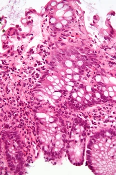
Appendicitis
Inflammation of the appendix, often requiring surgical removal.
Appendicitis is inflammation of the appendix, a common cause of sudden abdominal pain. It is primarily caused by a blockage of the hollow portion of the appendix, often by a calcified fecal deposit. If untreated, the appendix can rupture, leading to peritonitis and sepsis.
Quick Facts
- Field
- General surgery, emergency medicine
- Symptoms
- Periumbilical or right lower abdominal pain
- vomiting
- nausea
- decreased appetite
- high fever
- Complications
- Abdominal inflammation
- sepsis
- Gastrointestinal bleeding
- gastrointestinal abscess
- Diagnosis
- Based on symptoms
- medical imaging
- blood tests
- Differential
- Mesenteric adenitis
- cholecystitis
- psoas abscess
- abdominal aortic aneurysm
- Treatment
- Surgical removal of the appendix, antibiotics
- Frequency
- 11.6 million (2015)
- Deaths
- 50,100 (2015)
Facts from the source article.
Did You Know?
- If the appendix is retrocecal, deep pressure in the right lower quadrant may fail to elicit tenderness, a condition called silent appendix.
- The term 'appendicitis' was coined by American physician Reginald Heber Fitz in 1886.
Causes
Acute appendicitis appears to result from a primary obstruction of the appendix. Once blocked, the appendix fills with mucus and swells, increasing pressure within its lumen and walls. This pressure causes thrombosis and occlusion of small vessels and stasis of lymphatic flow, leading to ischemia and necrosis of the appendix. Bacteria then leak through the dying walls, forming pus (suppuration), and eventually the appendix may rupture, causing peritonitis, sepsis, and rarely death. The most common causative agents are calcified fecal deposits known as appendicoliths or fecaliths. Other causes include bezoars, foreign bodies, trauma, and lymphadenitis. Obstructing fecaliths occur more frequently in developed than in developing countries and are commonly associated with complicated appendicitis. Fecal stasis may play a role, as people with acute appendicitis have fewer bowel movements per week than healthy controls. Historically, diverticular disease and adenomatous polyps were unknown, and colon cancer was rare in communities where appendicitis was also rare, such as various African communities. Studies suggest that a transition to a Western diet lower in fiber is linked to rising frequencies of appendicitis and other colonic diseases in those communities. Low dietary fiber intake is thought to contribute to a right-sided fecal reservoir and reduced transit time.
Diagnosis
Diagnosis begins with a health history, symptom assessment, physical exam, and laboratory and imaging tests. Symptoms are categorized as typical or atypical. Typical appendicitis involves migratory right iliac fossa pain, nausea, anorexia (with or without vomiting), localized muscle stiffness, and mild fever. The classic progression is pain starting around the umbilicus and shifting to the right lower quadrant. Atypical histories lack this progression and may present with right lower quadrant pain as the initial symptom; irritation of the peritoneum can cause increased pain with movement or jolting. Physical exam findings become more apparent as the disease progresses and may include several signs: Aure-Rozanova's sign (increased pain on palpation in the right inferior lumbar triangle), Bartomier-Michelson's sign (increased pain in the right iliac region when lying on the left side), Dunphy's sign (increased right lower quadrant pain with coughing), Hamburger sign (patient refuses to eat, which is 80% sensitive for appendicitis), Kocher's sign (pain starting in the umbilical region and shifting to the right iliac region), Massouh's sign (a firm swish across the abdomen from the xiphoid process to the iliac fossae), and Obturator sign (pain with internal rotation of the flexed hip). When diagnosis is unclear, imaging such as ultrasound or CT scan is used; CT is more accurate, but ultrasound is often preferred first in children and pregnant women due to radiation concerns.
Management
Acute appendicitis is typically managed by surgery, though antibiotics are safe and effective for uncomplicated cases and may reduce overall complications. However, 51% of antibiotic-treated patients did not need an appendectomy after three years, while another study found 44% eventually had an appendectomy within ten years. Antibiotics are less effective if an appendicolith is present. The cost-effectiveness of surgery versus antibiotics is unclear. Antibiotics are recommended to prevent postoperative complications in emergency appendectomy and are effective when given before, during, or after surgery. Pain medications such as morphine do not appear to affect diagnostic accuracy and should be given early. The surgical removal of the appendix is called an appendectomy, which can be performed via open laparotomy (a single 2- to 3-inch incision in the lower right abdomen) or laparoscopic surgery. Laparoscopic appendectomy has several advantages over open appendectomy. A negative appendectomy—removal of a normal appendix with no inflammation on histopathology—occurs in about 13% of cases.
Prognosis
For most people, surgery leads to a quick recovery from appendicitis, though delays or the onset of peritonitis can bring complications. How long it takes to get better depends on factors like age, overall health, any complications, and how much alcohol someone drinks—usually between ten and twenty-eight days. A child around ten years old typically needs about three weeks to recover. Because peritonitis is a serious risk, anyone suspected of having appendicitis needs fast evaluation and treatment, and may require medical evacuation. In rare cases, when evacuation isn’t possible, appendectomies have been done outside a proper hospital. Typical acute appendicitis usually clears up quickly after the appendix is removed, and sometimes it goes away on its own; whether to schedule a later elective appendectomy to prevent a repeat episode is still debated. Atypical appendicitis, linked to suppurative cases, is harder to diagnose and more likely to cause problems even with early surgery. In both types, prompt diagnosis and removal of the appendix give the best outcome, with full recovery in two to four weeks. Death and serious complications are rare but can happen, especially if peritonitis isn’t treated. An appendicular lump can form if the appendix isn’t removed early, as the omentum and intestine stick to it; surgery during this phase is risky unless pus shows up on ultrasound or through fever and toxicity, so medical management is used instead. A rare complication after appendectomy is stump appendicitis, where the leftover stump becomes inflamed months or years later, and imaging like ultrasound can spot it.
More in Inflammatory Diseases of the Digestive System
Spotted an error? Know more?
Reader corrections go straight into our review queue. Suggest an edit · How this site is sourced