Streptococcus pneumoniae
A Gram-positive diplococcus causing pneumonia and other invasive infections.

Romina Camilli, Raoul JP Bonnal, Maria Del Grosso, Michele Iacono, Giorgio Corti · CC BY 2.0
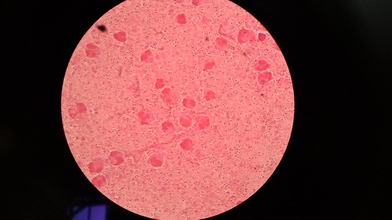
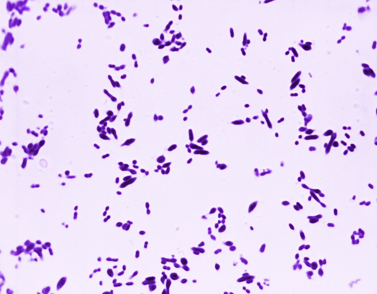
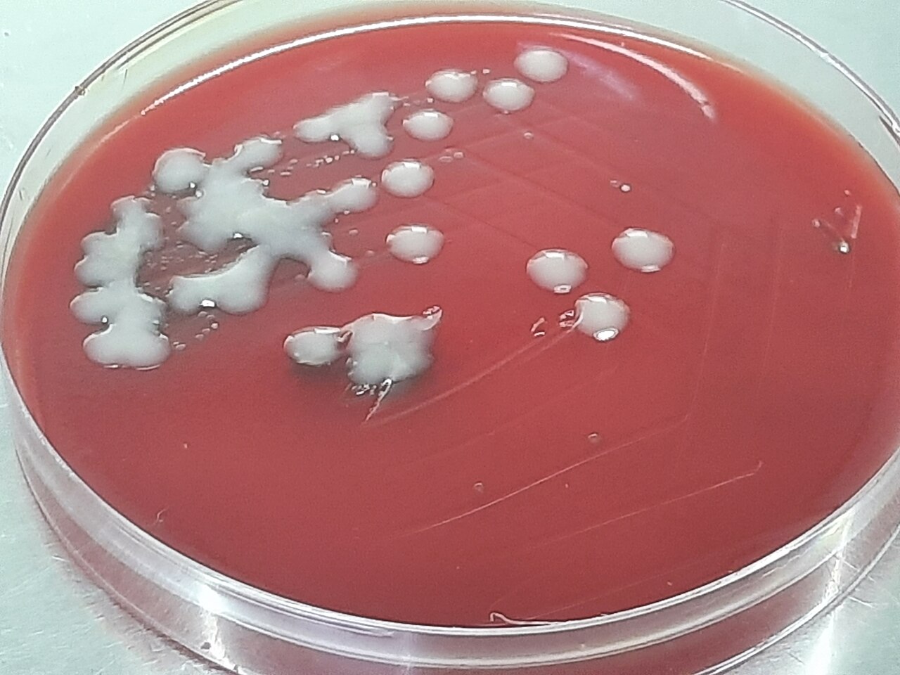
Streptococcus pneumoniae, also known as pneumococcus, is a Gram-positive, spherical, alpha-hemolytic bacterium that typically appears in pairs (diplococci). It is a significant human pathogen, recognized as a major cause of pneumonia in the late 19th century, and is the subject of many humoral immunity studies. The bacterium resides asymptomatically in healthy carriers, colonizing the respiratory tract, but can become pathogenic in susceptible individuals, causing a range of invasive diseases.
Quick Facts
- Genus
- Streptococcus
- Species
- pneumoniae
Facts from the source article.
Lore & Background
Streptococcus pneumoniae was first isolated simultaneously and independently in 1881 by U.S. Army physician George Sternberg and French chemist Louis Pasteur. It was termed Diplococcus pneumoniae in 1920 due to its appearance in Gram-stained sputum, and renamed Streptococcus pneumoniae in 1974 because of its similarity to streptococci. The organism played a central role in demonstrating that genetic material consists of DNA: in 1928, Frederick Griffith demonstrated transformation of life by turning harmless pneumococcus into a lethal form, and in 1944, Oswald Avery, Colin MacLeod, and Maclyn McCarty showed that the transforming factor was DNA, marking the birth of the molecular era of genetics.
Reader's Guide
Streptococcus pneumoniae is a leading cause of community-acquired pneumonia and meningitis in children and the elderly, and of sepsis in those infected with HIV. It also causes bronchitis, otitis media, sinusitis, and other invasive diseases. Its polysaccharide capsule acts as a key virulence factor, inhibiting phagocytosis, and more than 100 serotypes exist, varying in virulence and drug resistance. The bacterium undergoes phase variation between transparent and opaque colony phenotypes, allowing it to survive in different body systems. Natural transformation, induced by DNA-damaging agents, enables genetic exchange and likely aids in repairing oxidative DNA damage, contributing to virulence. The pneumococcal genome contains a core set of 1553 genes and a virulome of 154 genes. Understanding S. pneumoniae has been fundamental to molecular genetics and continues to inform vaccine development and antibiotic resistance research.
Did You Know?
- S. pneumoniae cells are usually found in pairs (diplococci) and do not form spores and are non-motile.
- The organism was first isolated in 1881 simultaneously by George Sternberg and Louis Pasteur.
- In 1944, Avery, MacLeod, and McCarty demonstrated that the transforming factor in Griffith's experiment was DNA, not protein.
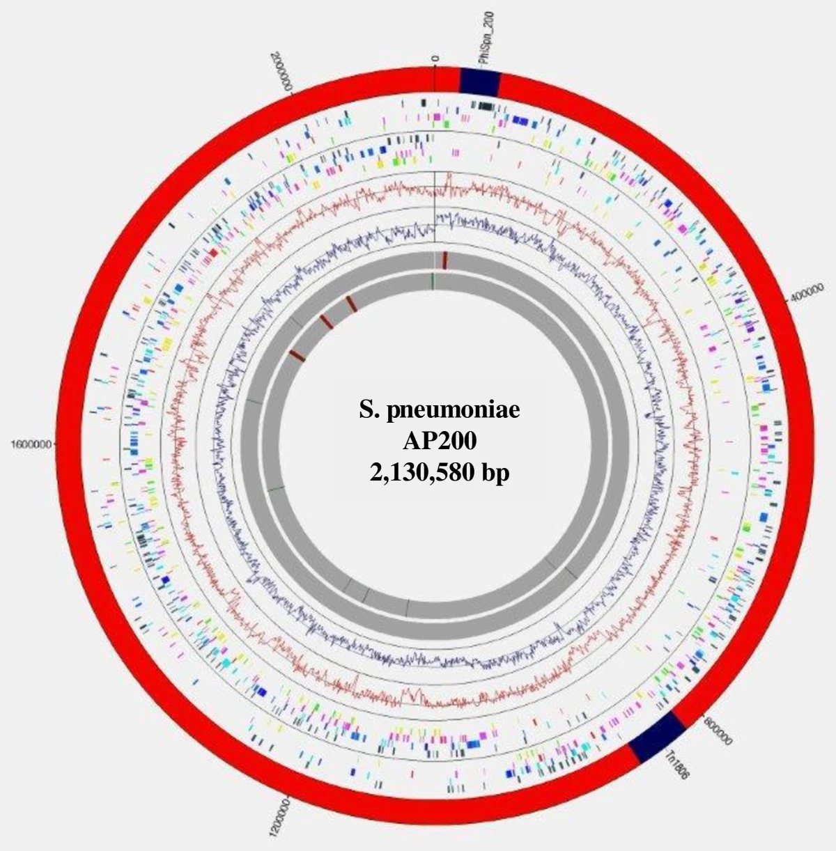
- The pneumococcal genome contains between 2.0 and 2.1 million base pairs and includes a variable number of lantibiotics depending on the strain.
Discovery & Taxonomic Journey
The story of pneumococcus begins in 1881, when two researchers working independently arrived at the same breakthrough. George Sternberg, a physician serving in the U.S. Army, and Louis Pasteur, the celebrated French chemist, each isolated the organism on their own. By 1886, it had earned the name pneumococcus, a nod to its established role as a principal cause of pneumonia. For decades it carried the binomial Diplococcus pneumoniae, a label reflecting the lancet-shaped pairs visible in Gram-stained sputum samples. It was not until 1974 that taxonomists reclassified it as Streptococcus pneumoniae, acknowledging its close kinship with other members of the streptococcal genus. Morphologically, the bacterium is a Gram-positive coccus that is alpha-hemolytic, non-motile, and incapable of forming spores. Its cells characteristically appear as diplococci, and the organism has been recognized as a major human pathogen since the closing decades of the nineteenth century, making it one of the earliest bacteria linked definitively to a specific clinical disease.
The Transformation Legacy
Few organisms have shaped the trajectory of modern genetics as profoundly as pneumococcus. In 1928, Frederick Griffith performed a landmark experiment in which he co-inoculated mice with live, non-virulent pneumococci alongside heat-killed virulent strains. The mice died, and live virulent bacteria were recovered, demonstrating that something from the dead cells had transformed the harmless ones into a lethal form. For sixteen years the identity of that transforming principle remained debated. In 1944, Oswald Avery, Colin MacLeod, and Maclyn McCarty conclusively showed it was DNA, not protein, overturning the prevailing assumption and inaugurating the molecular era of genetics. Beyond this historical milestone, natural transformation in S. pneumoniae is a biologically rich process requiring at least twenty-three dedicated genes and a specialized physiological state called competence. Competence is triggered by DNA-damaging agents including mitomycin C, fluoroquinolone antibiotics, and topoisomerase inhibitors. Research by Michod and colleagues suggests this mechanism functions as an adaptation for repairing oxidative DNA damage inflicted by host granulocytes, while Li and others demonstrated that an intact competence system is essential for both nasal colonization fitness and lung virulence.
The Capsule & Serotype Diversity
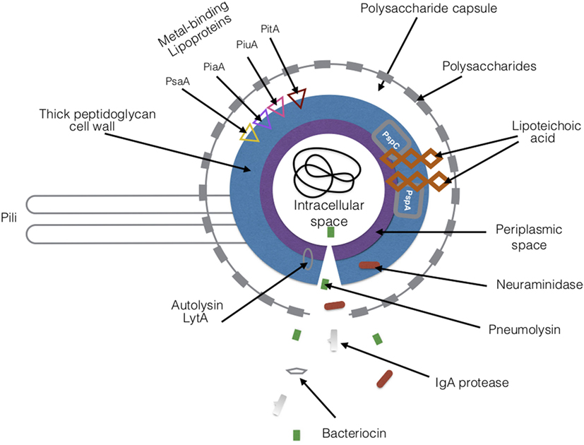
The defining weapon of encapsulated pneumococcus is its polysaccharide capsule, a thick outermost layer that sits atop the peptidoglycan cell wall. Chemically, it is a viscous, high-molecular-weight polymer built from repeating oligosaccharide units joined by covalent bonds. This structure serves as a formidable shield: by physically blocking granulocytes from reaching the cell wall beneath, the capsule effectively neutralizes phagocytosis, one of the host's primary innate defenses. The capsule is not a uniform structure across all strains. More than one hundred distinct serotypes have been catalogued, and they differ markedly in their chemical composition, the quantity of capsule they produce, their overall virulence, their geographic prevalence, and their susceptibility to antibiotic resistance. These serotype-specific variations are central to pathogenesis, dictating whether a given strain survives in the nasopharynx as a harmless commensal or invades deeper tissues to trigger life-threatening disease. In practical laboratory work, the capsule also aids identification: pneumococcus is distinguished from the similarly alpha-hemolytic viridans streptococci through its sensitivity to optochin and its characteristic lysis in bile, the so-called bile solubility test.
Clinical Spectrum & Transmission
In healthy carriers, pneumococcus lives quietly in the upper respiratory tract, sinuses, and nasal cavity without causing symptoms. Transmission occurs through direct person-to-person contact via respiratory droplets, and auto-inoculation from the upper airways can seed other body sites. The bacterium is also implicated in neonatal infections. Trouble arises when the host's immune defenses are compromised, as in the elderly, young children, or individuals living with HIV. In these vulnerable populations, the organism can breach local barriers and produce a remarkably wide array of invasive diseases. It is the leading cause of community-acquired pneumonia and bacterial meningitis in children and older adults, and a principal driver of sepsis among people infected with HIV. Beyond these headline conditions, pneumococcal infections encompass bronchitis, rhinitis, acute sinusitis, otitis media, conjunctivitis, osteomyelitis, septic arthritis, endocarditis, peritonitis, pericarditis, cellulitis, and brain abscess. Key virulence factors that enable this clinical breadth include the antiphagocytic capsule, the toxin pneumolysin, various adhesins, and immunogenic cell-wall components, all of which work in concert to overcome host defenses and establish invasive disease.
Gallery
Frequently Asked Questions
Who is Streptococcus pneumoniae?
Streptococcus pneumoniae, commonly called pneumococcus, is a Gram-positive, spherical bacterium that typically exists in pairs (diplococci) and shows alpha-hemolytic activity on blood agar. It was first isolated in 1881 and has since become one of the most studied human pathogens in microbiology.
What diseases does Streptococcus pneumoniae cause?
While it often lives harmlessly in the respiratory tract of healthy carriers, pneumococcus can invade vulnerable hosts and trigger pneumonia, meningitis, otitis media, and sepsis. Its polysaccharide capsule and other virulence factors help it evade immune defenses and spread from the nasopharynx to deeper tissues.
Why is Streptococcus pneumoniae historically significant?
Beyond being a leading cause of pneumonia and other invasive infections, pneumococcus was central to the landmark experiments that demonstrated DNA is the hereditary material. Its well-characterized transformation system made it a cornerstone organism for early molecular biology and humoral immunity research.
How many serotypes does Streptococcus pneumoniae have?
More than 100 distinct serotypes have been identified, each defined by a unique polysaccharide capsule structure. This serotype diversity is what makes broad-spectrum vaccine development so challenging, since antibodies against one capsule type do not confer protection against the others.
What does Streptococcus pneumoniae's genome look like?
The pneumococcus genome spans roughly 2.0 to 2.1 million base pairs, a relatively compact size for a bacterium of its ecological versatility. Its genetic toolkit includes numerous surface proteins and capsule biosynthesis genes that enable it to colonize and occasionally invade the human respiratory tract.
More in Infectious Agents And Pathogens 1-21
Spotted an error? Know more?
Reader corrections go straight into our review queue. Suggest an edit · How this site is sourced