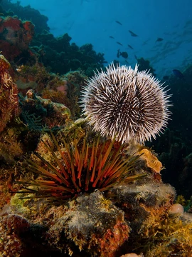
Sea urchin skeletogenesis
Larval skeleton formation in the sea urchin, critical yet transient.

Skeletogenesis in the sea urchin is a key morphogenetic event in embryonic development, involving the formation of a larval skeleton that is critical yet transient. The process has been most thoroughly studied in the species Strongylocentrotus purpuratus, an indirect developer whose larva must undergo metamorphosis to form the juvenile adult.
- Species studied
- Strongylocentrotus purpuratus
- Skeleton composition
- calcium carbonate (CaCO3) with 5% MgCO3
- Spicule crystallinity
- crystalline (confirmed by optical birefringence and X-ray diffraction)
- Spine dimensions
- 1-3 cm in length, 1-2 mm thick
- Time of skeletogenesis start
- 9-10 hours post fertilization (early blastula)
- Time of syncytial cable formation
- 13.5 hours post fertilization
- Time of pluteus stage
- 24 hours post fertilization
Lore & Background
Skeletogenesis begins in the early sea urchin blastula (9–10 hours post fertilization) when primary mesenchyme cells (PMCs), the sole descendants of the large micromere daughter cells, undergo an epithelial–mesenchymal transition and enter the blastocoel, forming a cell cluster at the vegetal pole. These PMCs, sometimes called the skeletogenic mesenchyme, exclusively produce the skeletal elements in a wild type embryo. Secondary mesenchyme cells (SMCs) have a skeletogenic potential, but signals from PMCs suppress this and direct SMCs into alternative pathways.
Once in the blastocoel, PMCs extend filopodia (250 nm in diameter, 25 μm long) that move randomly along the inner blastocoel wall. During the gastrula stage, after blastopore formation, PMCs localize in the prospective ventrolateral region and fuse into syncytial cables, forming the axis for calcium carbonate spicules (with 5% MgCO3) of the larval skeletal rods at 13.5 hours post fertilization. The spicules are crystalline. By the pluteus stage (24 hours post fertilization), extracellular matrix is abundant. The skeleton grows in size and complexity from gastrula to pluteus stages, and after metamorphosis, the larval skeleton is lost, though it gives rise to the juvenile's spines, which may be poisonous in some species.
Reader's Guide
The significance of sea urchin skeletogenesis lies in its role as a model for understanding morphogenetic events and cell interactions during development. The process is notable for the key interaction between PMCs and SMCs, where PMCs suppress the skeletogenic potential of SMCs, ensuring that only PMCs produce the larval skeleton. The molecular mechanisms involve PMC-specific gene products such as Msp30 (a sulfate cell-surface glycoprotein implicated in calcium uptake and deposition) and the spicule matrix proteins SM50, SM30, and PM27. SM50 and PM27 are nonglycosylated basic proteins, while SM30 is an acidic glycoprotein; their roles may involve nucleation or orientation of crystal growth. The msp130 gene shows complex spatial regulation within the PMC syncytium, and the ectoderm may help control skeletal morphogenesis by regulating PMC-specific gene expression.
Evolutionarily, comparative studies across distantly-related sea urchins and other echinoderms have revealed differences in spatiotemporal gene expression of transcription factors in the gene regulatory network for skeletogenic specification, yet striking similarities in signaling systems that position cells. Ancestral state reconstruction supports the homology of skeletogenic cells, suggesting this cell type arose before the divergence of cidaroids and euechinoids over 268 million years ago. The larval skeleton is critical yet transient, as it is lost after metamorphosis but gives rise to the juvenile's spines.
Did You Know?
- The larval skeleton is composed of calcium carbonate with 5% magnesium carbonate.
- Primary mesenchyme cells extend filopodia that are 250 nm in diameter and 25 μm long.
- The spicules are crystalline, as shown by optical birefringence and X-ray diffraction.
- The larval skeleton gives rise to the spines of the juvenile sea urchin, which may be poisonous in some species.
The Morphological Journey from Blastula to Juvenile
Skeletogenesis in the sea urchin is a dramatic yet fleeting chapter in embryonic development. Beginning roughly 9 to 10 hours after fertilization, primary mesenchyme cells detach from the apical epithelial layer of the early blastula and migrate into the blastocoel cavity. These cells extend remarkably thin filopodia—250 nanometers in diameter yet 25 micrometers long—that initially wander randomly across the inner blastocoel wall, forming and releasing attachments. By the gastrula stage, the cells settle into the ventrolateral region and fuse into syncytial cables that serve as scaffolds for calcium carbonate spicules, with a minor 5 percent magnesium carbonate component. Both optical birefringence and X-ray diffraction confirm the spicules are crystalline. At 24 hours, the pluteus larva displays abundant extracellular matrix alongside the syncytia. The skeleton grows in size and complexity from gastrula to pluteus, yet upon metamorphosis the larval skeleton is shed entirely. What remains is a paradox: a structure essential to larval life yet ultimately discarded, though the pluteus skeleton does seed the juvenile's spines, which can reach 1 to 3 centimeters in length and 1 to 2 millimeters in thickness, and in some species carry venom.
The Regulatory Dialogue Between Two Mesodermal Populations
A defining feature of sea urchin skeletogenesis is the regulatory interplay between primary mesenchyme cells and secondary mesenchyme cells. In a wild-type embryo, the larval skeleton is produced exclusively by PMCs, earning them the alternate designation of skeletogenic mesenchyme. Yet SMCs possess an inherent capacity for skeletal formation that remains unrealized under normal conditions. The PMCs actively suppress this latent skeletogenic potential in the SMCs, redirecting those cells toward alternative developmental fates. This interaction constitutes one of the principal regulatory relationships governing mesodermal cell behavior in the embryo. The PMC lineage itself traces back to the large micromere daughter cells, which undergo an epithelial-mesenchymal transition to liberate themselves from the apical layer. This EMT is the critical first step that frees the cells to enter the blastocoel and eventually form the syncytial cables scaffolding the larval skeleton. The entire process is thus governed not by a single cell type acting in isolation, but by a coordinated cellular dialogue in which one population actively constrains the developmental options available to the other.
Molecular Machinery of Spicule Biogenesis
The molecular toolkit driving spicule formation centers on a set of PMC-specific gene products whose precise roles remain only partially resolved. Msp30, a sulfate-containing cell-surface glycoprotein, has been linked to calcium uptake and deposition at the growing spicule. Three additional proteins—SM50, SM30, and PM27—constitute the organic spicule matrix. SM50 and PM27 appear to be structurally related, nonglycosylated, basic proteins, while SM30 is an acidic glycoprotein, suggesting functional diversity within the matrix. These matrix proteins are hypothesized to participate in nucleating crystal growth or orienting the crystalline lattice, though their exact mechanisms have not been fully elucidated. Beyond these structural components, the msp130 gene displays a complex spatial expression pattern within the PMC syncytium as skeletogenesis progresses, hinting at dynamic, region-specific regulation. Additionally, evidence suggests the ectoderm may exert influence over skeletal morphogenesis by modulating the expression of PMC-specific genes involved in spicule biogenesis, introducing an inter-tissue regulatory layer to the process.
Deep Evolutionary Roots of the Skeletogenic Program
The thorough characterization of skeletogenesis in Strongylocentrotus purpuratus has made this species a cornerstone for comparative evolutionary developmental biology. Researchers have extended these studies to distantly related sea urchins and other echinoderms to trace how the skeletogenic program evolved across the phylum. These comparative investigations reveal a striking pattern: while the signaling systems that position mesodermal cells within the embryo show remarkable conservation, the spatiotemporal expression of several transcription factors within the gene regulatory network has diverged substantially across the sea urchin clade. Differences in the timing of mesodermal ingression into the blastocoel and in the spatial patterns of transcription factor expression mark evolutionary shifts in the skeletogenic program. Nevertheless, ancestral state reconstruction of genes critical to skeletogenic cell specification points to the deep homology of this cell type. The evidence indicates that the skeletogenic mesenchyme lineage predates the split between cidaroids and euechinoids, a divergence that occurred over 268 million years ago, anchoring this developmental program in the ancient echinoderm past.
Frequently Asked Questions
What is sea urchin skeletogenesis?
It is the embryonic process in which a developing sea urchin larva builds its internal mineral skeleton, a structure that is essential for the larval stage but ultimately discarded once the animal metamorphoses into its adult form.
When does skeletogenesis begin in the sea urchin embryo?
The process kicks in roughly 9 to 10 hours after fertilization, while the embryo is still at the early blastula stage. By about 13.5 hours post-fertilization, a syncytial cable has formed to coordinate the crystallization work across the developing spicules.
What is the larval skeleton made of?
The skeleton consists primarily of calcium carbonate (CaCO3) with approximately 5% magnesium carbonate (MgCO3) incorporated into the lattice. The spicules are confirmed to be crystalline in structure, a finding verified through both optical birefringence and X-ray diffraction analyses.
Which sea urchin species is most commonly studied for skeletogenesis?
Strongylocentrotus purpuratus is the go-to model because it is an indirect developer, meaning its larva must undergo a full metamorphosis to become a juvenile adult. This makes the skeleton's formation and subsequent resorption especially well-defined and easy to track.
Why is sea urchin skeletogenesis considered a landmark in developmental biology?
It provides a clean, well-timed example of how a group of cells coordinates to produce a precisely shaped mineral structure from scratch, and because the skeleton is transient, it also reveals how an organism can dismantle its own architecture. The resulting larval spines reach 1–3 cm in length and 1–2 mm in thickness before being resorbed at metamorphosis.
More in Sea Urchins 1-24
Spotted an error? Know more?
Reader corrections go straight into our review queue. Suggest an edit · How this site is sourced