Cat-scratch disease
Infectious disease from cat scratches or bites.

Nephron · CC BY-SA 3.0
Cat-scratch disease (CSD) is an infectious disease most often resulting from a scratch or bite of a cat, caused by the bacterium Bartonella henselae. Symptoms typically include a painless bump or blister at the injury site, followed by painful, swollen lymph nodes, along with possible fatigue, headache, or fever. The disease affects approximately 1 in 10,000 people and is more common in children.
Quick Facts
- Field
- Infectious disease
- Symptoms
- Bump at the site of the bite or scratch, swollen and painful lymph nodes
- Complications
- Encephalopathy, parotitis, endocarditis, hepatitis
- Onset
- Within 14 days after infection
- Causes
- Bartonella henselae from a cat bite or scratch
- Diagnosis
- Based on symptoms, blood tests
- Differential
- Adenitis, brucellosis, lymphogranuloma venereum, lymphoma, sarcoidosis
- Treatment
- Supportive treatment, azithromycin
- Prognosis
- Generally good, recovery within 4 months
- Frequency
- 1 in 10,000 people
Facts from the source article.
Lore & Background
Cat-scratch disease commonly presents as tender, swollen lymph nodes near the site of inoculation, usually within 1–3 weeks. A painless vesicle or erythematous papule may form at the initial infection site. Most people also develop systemic symptoms such as malaise, decreased appetite, and aches. The disease usually resolves spontaneously within 2–4 weeks, though lymphadenopathy may persist for several months.
Reader's Guide
Cat-scratch disease is significant as a common zoonotic infection, particularly in children and young cat owners. While most cases are benign and self-limiting, rare complications can include serious neurologic or cardiac sequelae such as meningoencephalitis or endocarditis. Diagnosis is generally based on symptoms, with confirmation possible via blood tests or PCR. Treatment is primarily supportive, though antibiotics like azithromycin are recommended for severe disease or immunocompromised patients. Prevention focuses on flea control and hand washing after handling cats.
Did You Know?
- Young cats pose a greater risk than older cats for transmitting the disease.
- The bacterium Bartonella henselae is believed to be spread by the cat's saliva.
- Fleas serve as a vector for transmission of B. henselae among cats.
- The disease is specifically caused by Bartonella henselae from cats; transmission from dogs is not established as a cause of classic CSD.
The Tiny Invader and How It Travels
Bartonella henselae is a fastidious, intracellular, Gram-negative bacterium that has made the domestic cat its preferred host. Robert Debré identified the cat as the natural reservoir back in 1950, and the story of how the bug moves between species is genuinely intricate. The cat flea, Ctenocephalides felis, plays a central role: it carries viable B. henselae in its feces, and a cat can become infected when flea waste is scratched into the skin. Once established, kittens carry the organism in their bloodstream far more often than adult cats, making them the higher-risk transmitters. The jump to humans typically occurs when flea-contaminated scratch debris or infected saliva enters a wound. Ticks and even spiders have been implicated as occasional vectors, and researchers have flagged cryptic Bartonella exposure as a hidden occupational hazard for veterinarians. Although dog scratches or bites can rarely pass the infection along, the cat remains the overwhelmingly dominant source.
What the Body Experiences
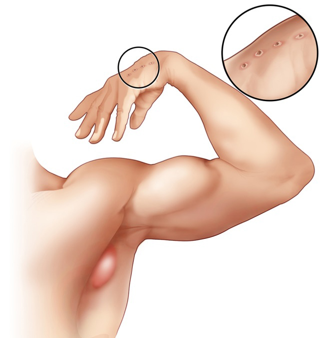
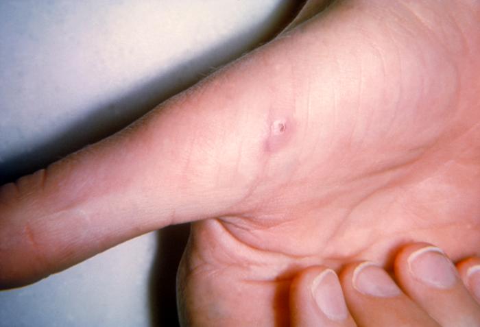
The first visible clue is often a small, non-painful bump or blister right where the scratch or bite landed. One to three weeks later, the regional lymph nodes nearest that site—most frequently in the armpit, along the arm, in the neck, or near the jaw—swell and become tender, usually on just one side. A vesicle or red papule may also appear at the original wound. Beyond the local signs, most patients develop a wave of systemic complaints: fatigue, loss of appetite, aching muscles and joints, headache, chills, and sometimes backache or abdominal pain. The timeline is variable; symptoms can surface as early as seven days after exposure or as late as two months. In the vast majority of cases the illness is benign and self-limiting, resolving within roughly a month even without intervention. Swollen nodes, however, can linger for several months after everything else has cleared. Full recovery generally takes about four months, though in some individuals it stretches toward a year. The condition touches roughly one in ten thousand people and hits children more often than adults.
Reading the Evidence: Diagnosis Under the Microscope
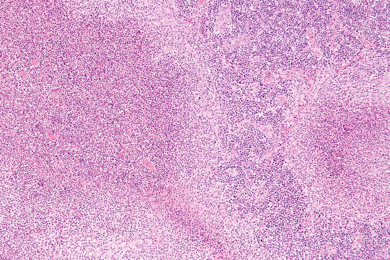
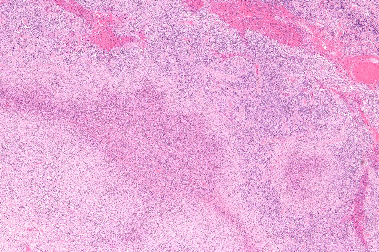
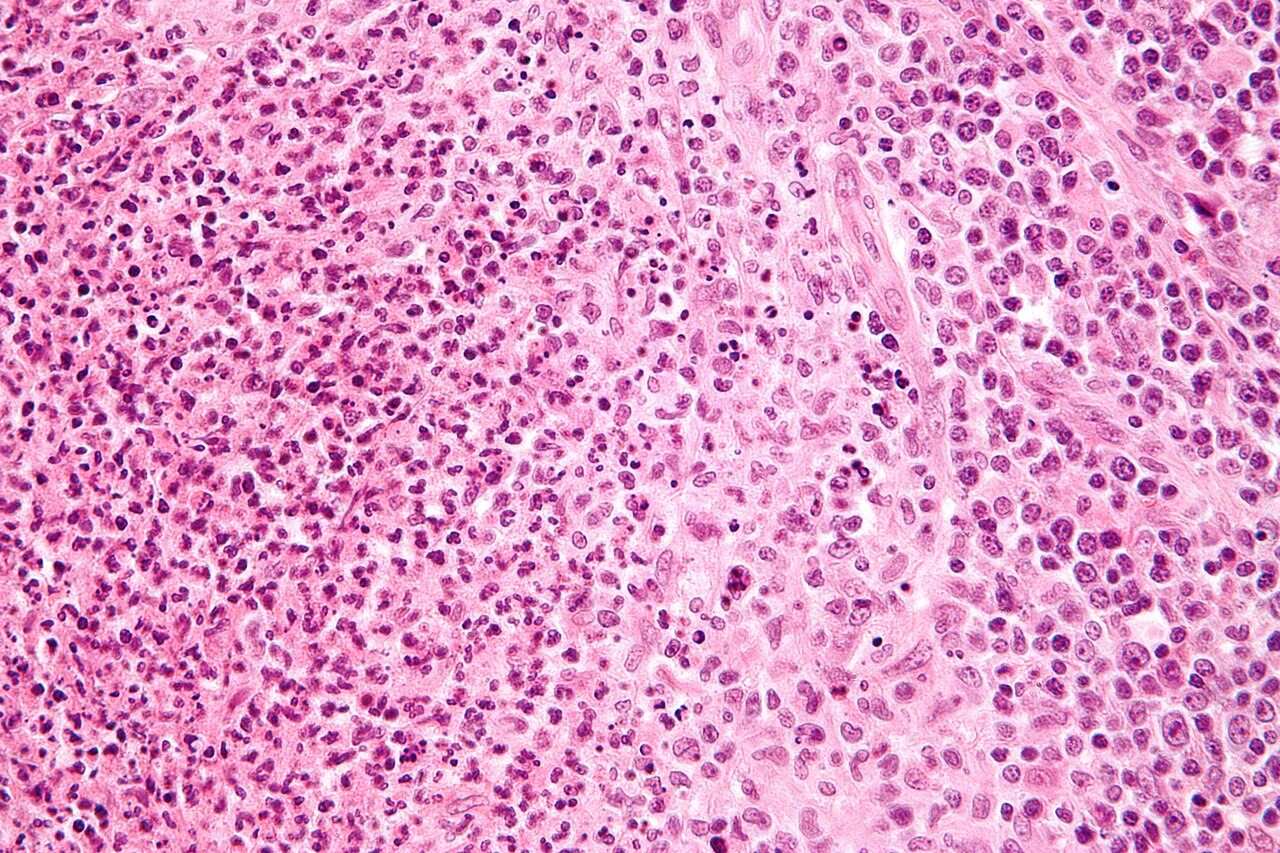
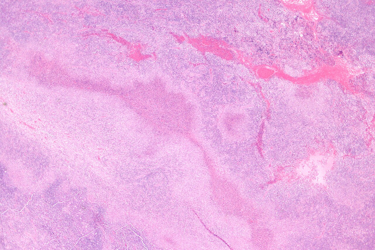
Because B. henselae is notoriously difficult to culture—incubation can take two to six weeks—clinicians lean heavily on indirect methods. Polymerase chain reaction is the most powerful confirmatory tool, with reported sensitivity ranging from 43 to 76 percent and, in at least one study, perfect specificity. The Warthin–Starry silver stain can reveal the organism in tissue, though interpreting the results is often tricky. Diagnosis in practice usually starts with the clinical picture and is backed up by blood tests. When a lymph node is biopsied, histology tells a distinctive story: the skin lesion shows a circumscribed patch of necrosis ringed by histiocytes, multinucleated giant cells, lymphocytes, and eosinophils. The node itself displays follicular hyperplasia with a central star-shaped zone of necrosis packed with neutrophils, surrounded by palisading histiocytes forming what pathologists call suppurative granulomas. Sinuses fill with monocytoid B cells. This classic pattern, while textbook, actually appears in only a minority of examined specimens.
When Things Go Wrong, and How to Stay Ahead
For most healthy individuals the infection clears on its own, and supportive care is the mainstay of management. Azithromycin is the preferred antibiotic when treatment is warranted—it is the only agent validated in a randomized controlled trial—and it is especially recommended for patients with severe disease or compromised immune systems. Still, in five to fourteen percent of cases the organism spreads to the liver, spleen, eye, or central nervous system. In rare but devastating scenarios, complications include meningoencephalitis, encephalopathy, seizures, and endocarditis, the last carrying a particularly grim mortality rate. Parinaud's oculoglandular syndrome, a granulomatous conjunctivitis paired with swelling of the node near the ear, is the most common eye-related manifestation, while optic neuritis represents an atypical presentation. Immunocompromised patients face additional threats such as bacillary angiomatosis and bacillary peliosis. Prevention centers on flea control, keeping cats indoors to limit outdoor flea exposure, and thorough hand-washing after handling cats or their feces, since infected cats show no symptoms at all.
Gallery
Frequently Asked Questions
Who is Cat-scratch disease?
Cat-scratch disease is a zoonotic infection triggered by the bacterium Bartonella henselae, usually introduced through a cat's scratch or bite. It is an uncommon condition, striking roughly 1 in 10,000 people, with children being the most frequently affected group.
What are Cat-scratch disease's powers/role?
After the initial wound, CSD produces a painless bump or small blister at the site, then drives painful swelling of nearby lymph nodes. It can also bring on fatigue, headaches, and low-grade fever as the immune system mounts its response.
Why is Cat-scratch disease important?
Although rarely severe, CSD is a common enough cat-related illness to be a notable concern for pet owners and pediatricians alike. Its higher prevalence in children makes it a frequently encountered diagnosis in young patients presenting with unexplained swollen glands.
What's Cat-scratch disease's origin?
The infection traces back to Bartonella henselae, a bacterium carried by cats—particularly kittens—and often maintained in their population through fleas. Human cases almost always begin when a cat's scratch or bite inoculates the bacteria directly into a wound.
More in Zoonoses 1-24
Spotted an error? Know more?
Reader corrections go straight into our review queue. Suggest an edit · How this site is sourced