Balantidium coli
The only ciliate protozoan known to infect humans.

The Other 95% · CC BY-SA 4.0
Balantidium coli is a parasitic ciliate alveolate responsible for the disease balantidiasis. It is the only ciliate known to cause illness in humans, though its primary reservoirs are domestic and wild pigs. The parasite also infects a range of other mammals—including cattle, camels, sheep, buffalo, and rodents—and, less commonly, some birds and marine mammals. Found worldwide, it is more prevalent in humid, moist environments. Human infections are most frequent in tropical and subtropical regions of Central and South America and Asia, though infection rates vary significantly from place to place.
First described in 1857 as *Paramecium coli*, it was moved to the genus *Balantidium* in 1863. In 2013, genetic analysis led to a proposal to reclassify it into its own genus, *Neobalantidium*, though this name is used interchangeably with *Balantioides*, a genus proposed in 1931.
**Morphology**
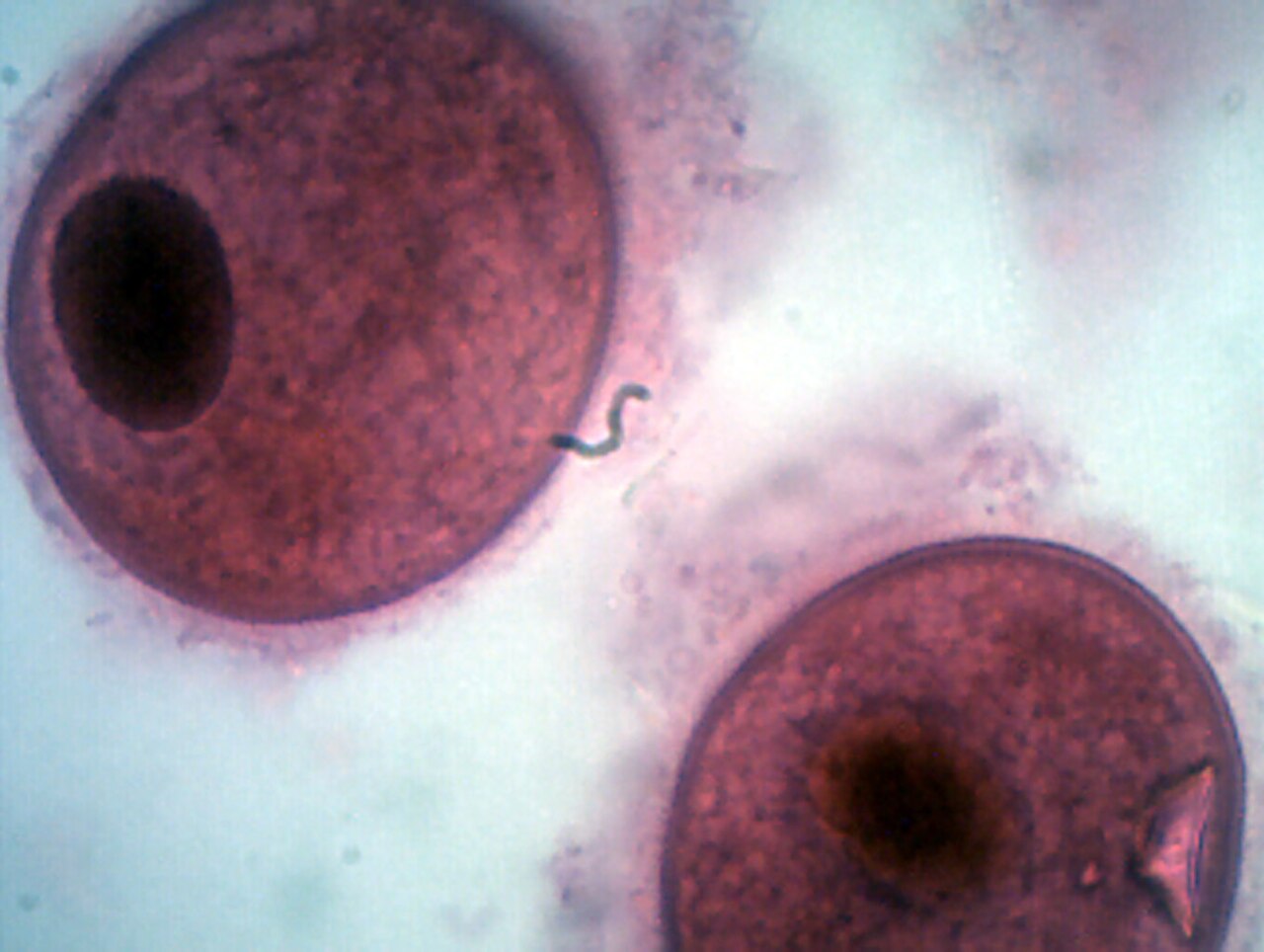
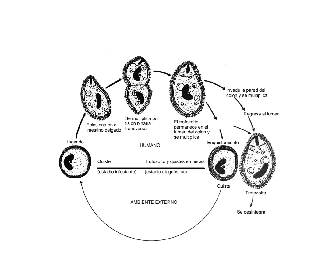
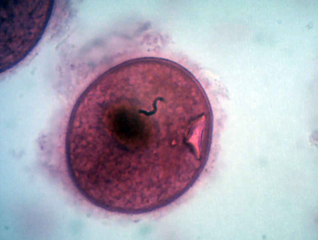
*B. coli* has two life stages: the trophozoite and the cyst. In the trophozoite stage, two nuclei are visible: a long, sausage-shaped macronucleus and a smaller, spherical micronucleus tucked beside it, often hidden. At the pointed front end, an opening called the peristome leads to the cytostome, or mouth. Cysts are smaller and rounder than trophozoites, with a tough wall made of one or two layers. Inside the cyst, only the macronucleus is usually visible, along with sometimes cilia and contractile vacuoles, though both nuclei are present because no nuclear multiplication occurs in this stage. Living trophozoites and cysts appear yellowish or greenish.
**Transmission**
Balantidiasis is a zoonotic disease. Humans acquire it through the fecal–oral route, most often from domestic pigs, which carry the parasite without symptoms. Contaminated water is the most common way the infection spreads.
**Role in Disease**
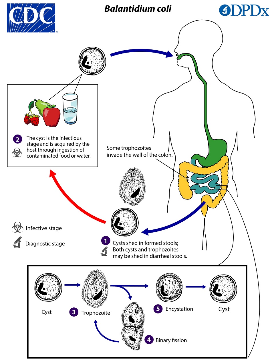
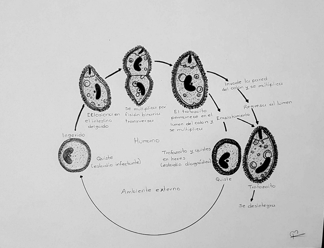
*B. coli* lives in the cecum and colon of humans, pigs, rats, and other mammals. It does not easily transfer between host species, as it needs time to adjust to the new host’s gut flora. Once adapted, it can become a serious pathogen, especially in humans. Trophozoites multiply and form cysts when feces dehydrate. Infection occurs when cysts are ingested, typically through contaminated food or water. In people with healthy immune systems, infection is possible but rarely causes severe gastrointestinal disease.
Quick Facts
- Taxon
- Balantidium coli
Facts from the source article.
Lore & Background
Balantidium coli was first described in 1857 as Paramecium coli before being transferred to the genus Balantidium in 1863. In 2013, genetic analysis comparing B. coli with other species in the genus led to a proposal to isolate it into its own genus Neobalantidium, though this name has been used interchangeably with the genus Balantioides proposed in 1931. The organism has two developmental stages: a trophozoite stage and a cyst stage. Trophozoites have two visible nuclei—a long, sausage-shaped macronucleus and a spherical micronucleus—and a peristome leading to the cytostome. Cysts are smaller, round, and have a tough wall; living trophozoites and cysts are yellowish or greenish in color.
Reader's Guide
Balantidium coli is significant as the only ciliated protozoan known to infect humans, causing the disease balantidiasis. Its primary reservoir is domestic and wild pigs, and transmission occurs via the fecal–oral route, most commonly through contaminated water. The infection is rare in humans (less than 1% of the population) and is most problematic in developing countries where water sources may be contaminated with swine or human feces. The cyst stage is infective due to its protective outer wall, surviving up to 10 days outside the host at room temperature and multiple weeks in feces, while the trophozoite survives only a few hours outside the host. Infection is more likely in malnourished individuals or those with compromised immune systems; acute disease can cause explosive diarrhea and colon perforation. The organism's ability to adapt to different hosts and its worldwide distribution, especially in tropical and subtropical regions, make it a notable zoonotic pathogen. Its classification remains under debate, with proposals to move it to the genus Neobalantidium or Balantioides.
Did You Know?
- Balantidium coli is the only member of the phylum Ciliophora known to be pathogenic to humans.
- The trophozoite form is unable to tolerate acids of pH 5 or lower, while the cyst can survive stomach acidity.
- In captive monkeys and primates, B. coli infection is more common than in their wild counterparts.
Taxonomic Odyssey & Classification
Balantidium coli holds a singular place in the animal kingdom as the sole ciliate alveolate within the phylum Ciliophora known to cause disease in humans. Its taxonomic identity, however, has been anything but stable. The organism was first formally described in 1857 under the name Paramecium coli, a classification that reflected the limited understanding of protozoan diversity at the time. Six years later, in 1863, researchers recognized enough distinguishing features to transfer it into the genus Balantidium. The story did not end there. In 2013, genetic comparisons between B. coli and its congeners prompted a proposal to elevate it into its own genus, Neobalantidium. This name has been used interchangeably with Balantioides, a genus proposed back in 1931. Despite these taxonomic shifts, the organism's biological significance remains constant: it is the only member of its entire phylum capable of pathogenicity in people, making its classification a matter of both scientific curiosity and public health relevance.
Morphology & Developmental Architecture
The organism exists in two distinct morphological forms that serve very different ecological purposes. The trophozoite is the active, feeding stage, characterized by a pointed anterior end bearing a peristome that opens into the cytostome, or mouth. Within its body, two nuclei are visible: a long, sausage-shaped macronucleus and a smaller spherical micronucleus that sits nestled beside it, often obscured by its larger neighbor. The cyst stage, by contrast, is a compact, rounded survival form encased in a tough wall composed of one or two layers. While the macronucleus and occasionally cilia and contractile vacuoles can be seen inside the cyst, both nuclei remain present even though nuclear division does not take place during this dormant phase. Cysts are noticeably smaller than their trophozoite counterparts. In living specimens, both stages display a yellowish or greenish hue, a subtle visual marker that distinguishes them from other intestinal organisms under the microscope.
Transmission Ecology & Geographic Reach
As a zoonotic pathogen, this protozoan primarily resides in domestic and wild pigs, where it causes no symptoms, yet it is capable of infecting a remarkably broad range of mammals including cattle, camels, sheep, buffalo, and various rodent species, with rare reports in certain birds and marine mammals. Human acquisition follows the fecal-oral route, most commonly through contaminated water supplies, though contaminated food and the use of infected manure as agricultural fertilizer also contribute to spread. The species enjoys a worldwide distribution but thrives particularly in humid, moist environments. Human cases cluster in tropical and subtropical regions of Central and South America and Asia, with the Philippines noted as a site of common occurrence. Despite its global presence, balantidiasis affects fewer than one percent of the human population. The cyst stage is the infective form, surviving up to ten days at room temperature and multiple weeks within feces, while the fragile trophozoite manages only a few hours outside a host or a few days in moist conditions.
Pathogenesis & Clinical Severity
Once ingested, the cyst must navigate the acidic gauntlet of the stomach. Its protective wall shields it from degradation, but at a pH below five the cyst is likely destroyed—a vulnerability that explains why malnourished individuals, whose gastric acidity is reduced, are at greater risk. Upon reaching the small intestine, trophozoites emerge and colonize the large intestine, feeding on resident flora. Some penetrate the colonic wall using proteolytic enzymes and multiply before returning to the lumen, where they may disintegrate or, triggered by dehydration of intestinal contents, encyst in the distal colon or within feces. In immunocompetent hosts, the protozoan often coexists without producing dysenteric symptoms. However, in those with compromised immunity or poor nutrition, acute disease can manifest as explosive diarrhea occurring as frequently as every twenty minutes. The most dangerous complication is colonic perforation, which can precipitate life-threatening emergencies.
Gallery
Frequently Asked Questions
Who is Balantidium coli?
Balantidium coli is a single-celled ciliate protozoan that dwells in the intestines of various mammals and is the sole member of its kind capable of making humans sick. It is most at home in domestic and wild pigs, but it can also colonize cattle, sheep, camels, buffalo, rodents, and, less commonly, some birds and marine mammals.
What is Balantidium coli's role in the zoonose lineup?
Its function in the zoonotic world is causing balantidiasis, an intestinal illness in humans acquired through the fecal-oral route, typically via contaminated water. It alternates between a motile trophozoite stage and a hardy cyst stage to persist in the environment and move between hosts.
How does Balantidium coli spread between hosts?
Transmission occurs when cysts shed in one animal's feces contaminate shared water or food and are then ingested by another pig, cow, or person. Humid tropical and subtropical settings—particularly across Central and South America and Asia—provide the moist conditions that make cross-species spread most common.
What's Balantidium coli's origin story?
The organism was first observed in 1857 and initially catalogued as Paramecium coli before being elevated to its own genus, Balantidium, in 1863. Taxonomists have continued to tweak its name—Balantioides appeared in 1931, and a 2013 proposal suggested Neobalantidium—so you will still encounter several synonyms in the literature.
Why does Balantidium coli matter in the zoonose canon?
It holds the unique distinction of being the only ciliate protozoan ever documented to cause disease in people, setting it apart from every other member of that broad group. Its worldwide distribution, especially in pig-heavy, humid regions, keeps it a persistent public-health concern in tropical and subtropical communities.
More in Zoonoses 1-24
Spotted an error? Know more?
Reader corrections go straight into our review queue. Suggest an edit · How this site is sourced