Scleritis
A serious inflammatory eye disease often linked to systemic disorders.
Scleritis is a serious inflammatory disease affecting the sclera, the white outer coating of the eye. It is often associated with systemic conditions such as granulomatosis with polyangiitis or rheumatoid arthritis and may be the first sign of connective tissue disease. The three types are diffuse scleritis (most common), nodular scleritis, and necrotizing scleritis (most severe).
Quick Facts
- Field
- Ophthalmology, Optometry
- Symptoms
- Eye redness
- pain
- photophobia
- tearing
- decrease in visual acuity
- Onset
- 30 - 60 years of age
- Risks
- Female gender
- Differential
- Episcleritis
- Treatment
- Non-steroidal anti-inflammatory drugs; Surgery (rarely needed)
- Frequency
- Uncommon
Facts from the source article.
Did You Know?
- Most of the time, scleritis is not caused by an infectious agent.
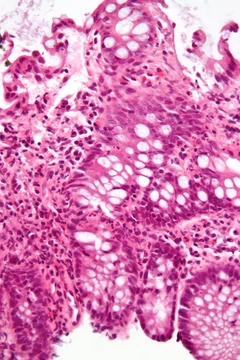
- Histopathological changes are those of a chronic granulomatous disorder with fibrinoid necrosis and infiltration by polymorphonuclear cells, lymphocytes, plasma cells, and macrophages.
- The granuloma is surrounded by multinucleated epitheloid giant cells and new vessels, some showing evidence of vasculitis.
Signs and symptoms
Symptoms include redness of the sclera and conjunctiva, sometimes with a purple hue, and severe ocular pain that may radiate to the temple or jaw, often described as deep or boring. Photophobia, tearing, and decreased visual acuity can occur, potentially leading to blindness. Secondary keratitis or uveitis may develop, with the most severe complications associated with necrotizing scleritis. Episcleritis pain is less severe, and unlike conjunctivitis, the redness does not move with gentle pressure.
Diagnosis
Scleritis is best detected by examining the sclera in daylight, with lid retraction to assess extent. Phenylephrine eye drops cause blanching in episcleritis but not in scleritis, aiding differentiation. Ancillary tests such as CT scans, MRIs, and ultrasonographies can be helpful but do not replace physical examination. Anterior scleritis accounts for about 98% of cases and is classified into non-necrotising (diffuse or nodular) and necrotising types; necrotising scleritis can occur with or without inflammation.
Treatment
For mild or moderate scleritis, doctors may prescribe NSAIDs like ibuprofen, indomethacin, or flurbiprofen to ease pain. If those don't work or the condition is posterior or necrotizing, systemic corticosteroids such as prednisolone are an option. Non-necrotizing cases can receive periocular steroid injections, but these are not used for necrotizing disease. When steroids alone fail, immunosuppressives or immunomodulators may be added. Surgery becomes necessary if the sclera thins excessively or perforates. Damaged corneal tissue can be repaired with a bandage contact lens or corneal glue.
More in Steroid-responsive Inflammatory Conditions
Spotted an error? Know more?
Reader corrections go straight into our review queue. Suggest an edit · How this site is sourced