Cerebral vasculitis
Inflammation of brain blood vessels causing stroke and varied symptoms.
Cerebral vasculitis, also termed cerebral angiitis, is inflammation of blood vessel walls within the brain and occasionally the spinal cord. It can affect capillaries, arterioles, venules, arteries, or veins, and reduced blood flow may lead to stroke. The condition produces a wide range of neurological symptoms, including headache, fatigue, joint pain, motor or sensory deficits, and altered cognition or behavior.
Quick Facts
- Incidence
- 2.4 cases per 1 million people
- Mortality
- 8-23%
- Risk of severe disability
- 25%
- Favorable response to therapy
- 75%
- Long-term remission with therapy
- 21-66%
Facts from the source article.
Did You Know?
- The most frequent presenting signs and symptoms are focal neurological deficits (63% of cases), headaches (51%), and cognitive impairment (41%).
- Spinal cord involvement is rare, especially when it does not also affect the brain.
- 10% of cases have associated bleeding in the brain.
Causes
Primary angiitis of the central nervous system (PACNS) has no identified secondary cause and its exact mechanism is unknown. Secondary cerebral vasculitis may arise from infections such as varicella zoster virus, systemic autoimmune diseases like systemic lupus erythematosus and rheumatoid arthritis, medications and drugs including amphetamine, cocaine, and heroin, certain cancers such as lymphomas, leukemia, and lung cancer, or other systemic vasculitides like granulomatosis with polyangiitis, polyarteritis nodosa, or Behçet's disease. Conditions that mimic cerebral vasculitis include fibromuscular dysplasia and thrombotic thrombocytopenic purpura. Older age, cognitive impairment at diagnosis, cerebral infarctions on imaging, spinal cord involvement, diagnostic delays, and medium or large vessel involvement are associated with poor prognosis and increased mortality.
Pathophysiology
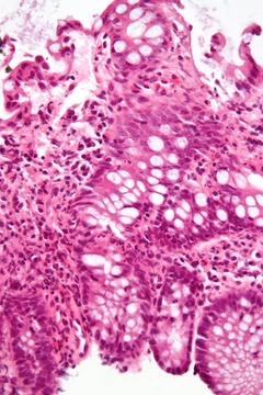
Three main histologic types of blood vessel inflammation are seen in primary CNS vasculitis. Granulomatous vasculitis, found in 32-61% of cases, features well-formed granulomas with multinucleated giant cells. Lymphocytic vasculitis, present in 24-79% of cases, involves lymphocytic infiltration without granulomas and is linked to less disability and mortality. Necrotizing vasculitis, occurring in 14-42% of cases, shows transmural fibrinoid necrosis and carries a high risk of intracerebral hemorrhage.
Diagnosis
Diagnosis of primary CNS vasculitis relies on brain imaging and biopsy of affected blood vessels, with biopsy being the most definitive modality. However, brain biopsy has a low yield, with 30-50% of biopsies normal in suspected cases due to irregular vessel involvement or inability to biopsy larger vessels safely. Digital subtraction angiography is the preferred imaging method, offering greater specificity than magnetic resonance angiography or CT angiography; typical findings include smooth wall segmental stenosis of multiple arteries, sometimes with post-stenotic dilation or bleeding. In small vessel disease, angiography is usually normal, and European guidelines recommend biopsy when angiography is normal but small vessel disease is suspected. For medium to large vessel disease with characteristic angiographic findings, biopsy is typically not required. Biopsy and angiography show low concordance: in one meta-analysis, digital subtraction angiography was positive in only 33% of biopsy-confirmed cases, and biopsy was positive in only 8% of angiographically confirmed cases. High-resolution MRI may show vessel wall hyperenhancement, multiple infarcts, leptomeningeal enhancement, and intracranial hemorrhages; a normal MRI makes the diagnosis unlikely. Inflammatory markers such as C-reactive protein and erythrocyte sedimentation rate are usually normal, while cerebrospinal fluid analysis shows a nonspecific pattern with mildly elevated leukocytes and increased protein, especially in small vessel disease.
Treatment
Glucocorticoids and cyclophosphamide are the mainstays of treatment for primary CNS vasculitis, with limited evidence for mycophenolate mofetil. Therapy typically involves an induction phase followed by at least two years of maintenance therapy, which is associated with lower deaths, lower relapse risk, and less disability. Rituximab or tocilizumab may be used for refractory disease, and European guidelines recommend adding aspirin for medium or large vessel involvement.
More in Steroid-responsive Inflammatory Conditions
Spotted an error? Know more?
Reader corrections go straight into our review queue. Suggest an edit · How this site is sourced