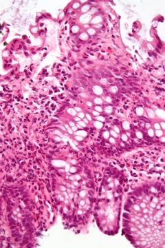
Chronic sclerosing sialadenitis
Benign salivary gland inflammation mimicking malignancy, linked to IgG4-related disease.
Chronic sclerosing sialadenitis is a rare, benign inflammatory condition of the salivary gland that presents as hard, indurated masses clinically indistinguishable from neoplasms. It is now regarded as a manifestation of IgG4-related disease.
Quick Facts
- Field
- Dentistry
Facts from the source article.
Did You Know?
- Standard therapy is glandular sialoadenectomy, associated with fairly low operative morbidity.
- Steroid administration can shrink the inflammatory lesion and reduce serum IgG4 values.
- Steroids may be useful in younger patients or those who refuse surgery.
Presentation
The inflammatory lesions may be unilateral or bilateral, predominantly involving the submandibular gland but also occurring in other major and minor salivary glands, including the parotid. Salivary gland tumors are rare, with salivary malignancies accounting for 3–5% of head and neck cancers; however, 20–25% of parotid, 35–40% of submandibular, and over 90% of sublingual gland tumors are malignant. This diagnostic challenge arises because Küttner's tumor, though benign, mimics malignancy clinically. The swollen masses are generally painful, and surgical resection is advised upon suspicion of malignancy; definitive diagnosis is made only by post-surgical histopathology.
Histological features
Histopathological features include heavy infiltration by lymphocytes and plasma cells (lymphoplasmacytic infiltrate), reactive lymphoid follicles without atypical lymphoid cells, atrophy and loss of acini, periductal fibrosis encasing glandular ducts in thick fibrous tissue, and eventual periductal and interlobular sclerosis.
Pathogenesis
The cause is not well understood. Proposed factors include sialolithiasis (hard salivary calculus formation), ductal abnormalities leading to retained secretions, and an autoimmune cause supported by the presence of abundant IgG4-associated plasma cells and increased serum IgG4. Sialoliths are observed in an appreciable proportion of submandibular cases but may be absent in many. The condition is primarily seen in adults aged 40–70 years, though a case with prominent immunopathological features was described in an 11-year-old boy in Brazil in 2012.
Diagnosis
Pre-operative diagnosis is difficult, and the condition remains under-reported. Surgeons increasingly rely on ultrasonography and fine-needle aspiration cytology (FNAC) for presumptive diagnosis. Ultrasonography shows a diffuse, heterogeneous zone of echo-shadows. FNAC reveals paucicellularity with scattered tubular ducts against a background of lymphoplasmacytic infiltration and fibrous depositions; findings may be nonspecific and require adjunct clinical and ultrasonographic correlation. MRI has been tried but results are inconclusive.
More in Steroid-responsive Inflammatory Conditions
Spotted an error? Know more?
Reader corrections go straight into our review queue. Suggest an edit · How this site is sourced