Autoimmune pancreatitis
A rare pancreatitis that mimics cancer but responds to steroids.
Autoimmune pancreatitis (AIP) is a rare form of chronic pancreatitis that often presents as a pancreatic mass, making it difficult to distinguish from pancreatic carcinoma. Its clinical importance lies in the fact that it responds well to corticosteroid treatment, whereas unnecessary pancreatic surgery carries substantial mortality and morbidity. Two types exist: Type 1, a manifestation of IgG4-related disease, and Type 2, which primarily affects the pancreas.
Quick Facts
- Field
- Gastroenterology
- Symptoms
- Painless jaundice, pancreatic mass
- Types
- Type 1 and Type 2
- Causes
- IgG4-related disease
- Diagnosis
- Biopsy
- imaging
- serology
- Differential
- Pancreatic cancer
- Treatment
- Corticosteroids (first line)
- azathioprine
- rituximab
Facts from the source article.
Did You Know?
- Type 1 AIP is more common in East Asia, whereas type 2 is relatively more common in the US and Europe.
- Terms frequently encountered for AIP include lymphoplasmacytic sclerosing pancreatitis and non-alcoholic duct destructive chronic pancreatitis.
- Some of the earliest cases were reported as pancreatic pseudotumor or pseudolymphoma.
Signs and symptoms
Two-thirds of patients present with either painless jaundice due to bile duct obstruction or a mass in the head of the pancreas, mimicking carcinoma. Type 1 AIP typically occurs in a 60–70-year-old male with painless jaundice; imaging may show a mass or diffuse pancreatic enlargement, and strictures in the pancreatic duct can occur. Rarely, Type 1 presents with acute pancreatitis. Systemic involvement occurs in at least half of Type 1 cases, most commonly cholangitis (up to 80 percent), along with possible inflammation in salivary glands, lungs (pulmonary fibrosis and nodules), mediastinal fibrosis, retroperitoneal fibrosis, or tubulointerstitial nephritis. Features include scleral icterus and painless jaundice without acute pancreatitis attacks, mild symptoms such as minimal weight loss or nausea, elevated serum gamma globulins, IgG or IgG4, and autoantibodies such as ANA, anti-lactoferrin, anti-carbonic anhydrase II, and rheumatoid factor. CT often shows a diffusely enlarged sausage-shaped pancreas, and ERCP reveals diffuse irregular narrowing of the main pancreatic duct and stenosis of the intrapancreatic bile duct. Pancreatic calcification or cyst formation is rare, and the condition shows marked responsiveness to corticosteroids.
Histopathology
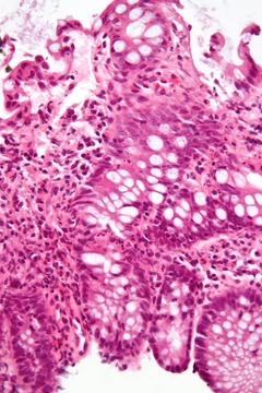
Histopathologic examination reveals a characteristic lymphoplasmacytic infiltrate of CD4- or CD8-positive lymphocytes and IgG4-positive plasma cells, with interstitial fibrosis and acinar cell atrophy in later stages. Early stages show a cuff of lymphoplasma cells surrounding the ducts and diffuse infiltration in the lobular parenchyma, though localization and degree of duct wall infiltration vary. Histopathology remains the primary method for differentiating AIP from acute and chronic pancreatitis, lymphoma, and cancer. Fine needle aspiration can yield a diagnosis if adequate tissue is obtained, with lymphoplasmacytic infiltration of lobules as the key finding; rarely, granulomatous reaction may be observed. A cytologic smear primarily composed of acini rich in chronic inflammatory cells with rare ductal epithelial cells lacking atypia favors AIP, though sensitivity and specificity for differentiating from neoplasia are unknown. In systemic AIP, similar pathologic features appear in other organs. The development of IgG4 antibodies recognizing an epitope on pancreatic acinar cells is considered an important factor in pathophysiology, though the auto-antigens involved remain obscure; an earlier suggestion that human ubiquitin-protein ligase E3 component n-recognin 2 (UBR2) was an antigen is likely an artifact.
Diagnosis
The International Consensus Diagnostic Criteria (ICDC) for AIP emphasize five cardinal features: imaging appearance of pancreatic parenchyma and duct, serum IgG4 level, other organ involvement with IgG4-related disease, pancreatic histology, and response to steroid therapy. The Japanese Pancreas Society criteria require pancreatic imaging showing diffuse narrowing of the main pancreatic duct with irregular wall over more than one-third of the pancreas, plus either laboratory data showing elevated serum gamma globulin or IgG or autoantibodies, or histopathologic findings of fibrotic changes with lymphocyte and plasma cell infiltrate. CT findings include a diffusely enlarged hypodense pancreas or a focal mass that may be mistaken for malignancy, with a low-density capsule-like rim possibly corresponding to peripancreatic inflammation. MRI shows diffusely decreased signal intensity and delayed enhancement on dynamic scanning. ERCP reveals segmental or diffuse irregular narrowing of the main pancreatic duct, often with an extrinsic-appearing stricture of the distal bile duct; changes similar to primary sclerosing cholangitis may occur in extrapancreatic bile ducts. EUS findings include a diffusely swollen and hypoechoic pancreas or a solitary focal irregular mass, but the role of EUS-FNA in AIP diagnosis remains unclear.
Treatment
AIP often completely resolves with steroid treatment. Failure to differentiate AIP from malignancy may lead to unnecessary pancreatic resection; a trial of steroid therapy could prevent a Whipple procedure or complete pancreatectomy for this benign disease. If relapse occurs after corticosteroids or they are not tolerated, immunomodulators such as azathioprine and 6-mercaptopurine may extend remission. If corticosteroids and immunomodulators are insufficient, rituximab can induce and maintain remission.
More in Steroid-responsive Inflammatory Conditions
Spotted an error? Know more?
Reader corrections go straight into our review queue. Suggest an edit · How this site is sourced