Autoimmune hepatitis
Chronic liver inflammation driven by autoimmune attack on hepatocytes.
Autoimmune hepatitis is a chronic autoimmune disease in which the immune system attacks liver cells, causing inflammation. It was formerly known as lupoid hepatitis, plasma cell hepatitis, or autoimmune chronic active hepatitis. The disease is notable for its bimodal age distribution and strong female predominance.
Quick Facts
- Symptoms
- Often asymptomatic
- fatigue
- right upper abdominal pain
- anorexia
- nausea
- jaundice
- joint pain
- rash
- Complications
- Chronic liver disease, cirrhosis
- Onset
- Bimodal presentation: 10-20 years of age, 40-50 years of age
- Duration
- Lifelong
- Types
- Type 1
- type 2
- seronegative
- Causes
- Genetic predisposition with environmental trigger
- Risks
- Female gender, additional autoimmune disease
- Differential
- Primary biliary cholangitis; Primary sclerosing cholangitis
Facts from the source article.
Did You Know?
- Autoimmune hepatitis was originally described in the early 1950s.
- African Americans appear to present with more aggressive disease and worse outcomes.
- The disease was previously called 'lupoid' hepatitis due to its association with systemic lupus erythematosus.
Signs and symptoms
Autoimmune hepatitis may be asymptomatic in 12–35% of cases or present with signs of chronic liver disease, acute hepatitis, or fulminant hepatic failure. Common nonspecific symptoms include fatigue, malaise, weight loss, right upper quadrant pain, nausea, jaundice, and joint pain affecting small joints. Amenorrhoea is frequent in women. Physical examination may be normal or reveal chronic liver disease signs. Many individuals are diagnosed after incidental laboratory abnormalities, such as elevated transaminases, while alkaline phosphatase and bilirubin are usually normal. The condition can overlap with type 1 diabetes mellitus, ulcerative colitis, lupus, celiac disease, vasculitis, and autoimmune thyroiditis.
Cause
Autoimmune hepatitis likely results from a mix of inherited risk, a trigger like a virus, drug, herb, or vaccine, and a breakdown in immune regulation, causing ongoing liver cell inflammation and scarring. The exact cause is unknown. About 60% of patients show signs of chronic hepatitis without viral markers. Anti-smooth muscle antibodies are a strong marker. Early, severe cases often link to HLA-DR3, while later-onset disease links to HLA-DR4.
Diagnosis
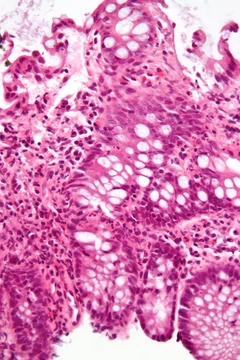
Diagnosis combines clinical, laboratory, and histological findings after excluding viral, hereditary, metabolic, cholestatic, and drug-induced liver diseases. Liver biopsy is required to obtain tissue for histological examination. Useful blood tests include antinuclear antibody (ANA), anti-smooth muscle antibody (SMA), anti-liver kidney microsomal antibodies (LKM-1, LKM-2, LKM-3), anti-soluble liver antigen (SLA), liver–pancreas antigen (LP), and anti-mitochondrial antibody (AMA); the latter is more suggestive of primary biliary cholangitis. Increased immunoglobulin G and hypergammaglobulinemia are also of diagnostic value. Histological findings include portal mononuclear cell infiltrate invading the portal triad boundary, interface hepatitis sparing the biliary tree, plasma cell infiltrate, hepatocyte rosettes, multinucleated giant cells, and varying degrees of fibrosis. The Internal Autoimmune Hepatitis Group developed a standardized scoring system for population studies, and a simplified scoring system for clinical use incorporates autoantibody titers, IgG levels, histology, and exclusion of viral hepatitis. Based on autoantibodies, three subtypes are recognized but have no distinct clinical presentations. Type 1 is positive for ANA, anti-smooth muscle antibody (in 65% of people), anti-actin antibodies, anti-mitochondrial antibodies (rare except in overlap syndromes), anti-soluble liver antigen/liver pancreas antibody (in 20%), and anti-double stranded DNA (in 30%).
Treatment
Treatment choice depends on symptom severity, elevation of liver enzymes and antibodies, liver biopsy findings, and tolerance of side effects. Asymptomatic patients with normal enzymes, antibodies, and non-inflammatory biopsies generally do not require treatment due to low risk of progression. For symptomatic individuals with interface hepatitis and necrosis on biopsy, treatment is recommended, especially in younger patients. Some authorities advise treating all diagnosed patients even if asymptomatic. The mainstay is immunosuppressive glucocorticoids such as prednisone during acute episodes, achieving symptom resolution in 60–80% of cases, though many relapse. For moderate-to-severe disease in those intolerant to glucocorticoids, lower-dose prednisone monotherapy or combination with azathioprine is an alternative. Budesonide may be more effective than prednisone for inducing remission, but evidence is limited. Non-responders to glucocorticoids and azathioprine may receive mycophenolate, ciclosporin, tacrolimus, or methotrexate. Cirrhosis develops in 7–40% of treated patients, with highest risk in those with incomplete response, treatment failure, or multiple relapses. Once cirrhosis occurs, management is standard regardless of etiology. Liver transplantation is standard for fulminant liver failure or disease progression despite multiple therapies. Many patients remain on long-term immunosuppression for life; common practice is to discontinue after two or more years of normalized transaminases and IgG, but approximately 90% relapse after stopping, leading some specialists to advocate permanent therapy.
More in Steroid-responsive Inflammatory Conditions
Spotted an error? Know more?
Reader corrections go straight into our review queue. Suggest an edit · How this site is sourced