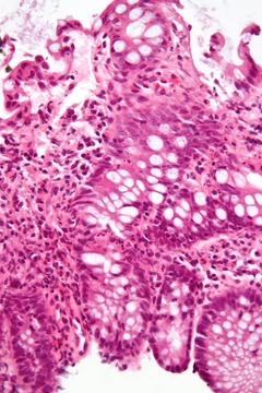
Atopic dermatitis
Chronic inflammatory skin condition with itchy, red, cracked skin.
Atopic dermatitis (AD), also known as atopic eczema, is a chronic inflammatory skin condition characterized by itchy, red, swollen, and cracked skin that may ooze clear fluid and thicken over time. It affects about 20% of people at some point in life, is more common in younger children, and females are affected slightly more often than males. The condition often begins in childhood and can vary in severity over the years, with many people outgrowing it.
Quick Facts
- Field
- Dermatology, Clinical Immunology and Allergy
- Symptoms
- Itchy
- red
- swollen
- cracked skin
- Complications
- Skin infections
- hay fever
- asthma
- Onset
- Childhood
- Risks
- Family history
- living in a city
- dry climate
- Diagnosis
- Based on symptoms after ruling out other possible causes
- Differential
- Contact dermatitis
- psoriasis
- seborrheic dermatitis
- Frequency
- ~20% at some time
Facts from the source article.
Did You Know?
- Eczema often starts on the cheeks and outer limbs in infants and later settles in skin folds such as behind the knees, elbows, neck, wrists, and under the buttock folds.
- Colonization by Staphylococcus aureus is prevalent in those with atopic dermatitis, and the bacterium exploits skin barrier abnormalities to trigger cytokine expression, aggravating the condition.
- The UK Diagnostic Criteria, based on the work of Hanifin and Rajka, is the most widely validated set of criteria for diagnosing atopic dermatitis.
Causes
The exact cause of atopic dermatitis is unknown, but it is considered a multifactorial disease likely arising from a combination of environmental, immunologic, bacterial, and genetic factors. A null filaggrin (FLG) gene is increasingly thought to play a significant role, though further study is needed. Rates of AD in the US and UK have increased three- to sixfold since 1970, and people who migrate from developing nations before age 4 to industrialized nations experience a dramatic rise in risk, especially when living in urban areas. Early life industrial exposures may cause AD; chemicals such as diisocyanates and xylene prevent skin bacteria from producing ceramide-sphingolipid lipids, and early life deficiency in these lipids predicts later development of AD. These chemicals also activate the itch receptor TRPA1. Industrial manufacturing and use of xylene and diisocyanates greatly increased starting in 1970, expanding average exposure. Low humidity and low temperature increase prevalence and risk of flare-ups. Genes contributing to AD include those responsible for immune response (e.g., TH2 cytokine and JAK-STAT pathway genes) and skin barrier (e.g., filaggrin, claudin-1, loricrin). Up to 80% of people with AD have elevated total or allergen-specific IgE levels.
Pathophysiology
Excessive type 2 inflammation underlies the pathophysiology of atopic dermatitis. Disruption of the epidermal barrier allows allergens to penetrate deeper skin layers, activating epidermal inflammatory dendritic and innate lymphoid cells, which attract Th2 CD4+ helper T cells to the skin. This dysregulated Th2 inflammatory response leads to eczematous lesions. Activated Th2 cells release inflammatory cytokines including IL-4, IL-13, and IL-31, which activate downstream Janus kinase (Jak) pathways, leading to inflammation and activation of plasma cells and B lymphocytes that release antigen-specific IgE. Other CD4+ helper T-cell pathways involved include Th1, Th17, and Th22; specific pathways are more commonly activated in certain ethnic groups, such as Th2 and Th17 in Asian people. Mutations in the filaggrin gene FLG cause impairment in the skin barrier. Filaggrin produced by keratinocytes stimulates release of moisturizing factors and lipid matrix material that contribute to skin barrier adhesion. Loss-of-function FLG mutations cause loss of this lipid matrix and moisturizing factors, leading to transdermal water loss, dry skin, and antigen penetration. Filaggrin mutations also decrease natural antimicrobial peptides, disrupting skin flora and promoting Staphylococcus aureus overgrowth.
Treatments
No cure for atopic dermatitis is known, but treatments can reduce severity and frequency of flares. The most commonly used topical treatments are corticosteroids to control flare-ups and moisturisers to maintain control. Daily basic care stabilizes skin barrier function; moisturisers can improve skin comfort and reduce flares, with all types equally effective. Emollient bath additives provide no additional benefit. Topical corticosteroids are effective; newer second-generation ones such as fluticasone propionate and mometasone furoate are more effective and safer than older ones. Strong and moderate corticosteroids are more effective than weaker ones, and applying once daily is as effective as more frequent application. Topical calcineurin inhibitors like tacrolimus and pimecrolimus are also first-line therapies, effective and safe. Crisaborole, a PDE-4 inhibitor, is effective and safe for mild-to-moderate AD. Ruxolitinib, a Janus kinase inhibitor, has uncertain efficacy and safety. Difamilast, a PDE4 inhibitor, was approved for medical use in Japan in September 2021 and in the United States in February 2026. More severe cases may need systemic medicines such as ciclosporin, methotrexate, dupilumab, or baricitinib.
Impact
Atopic dermatitis significantly impairs quality of life, with effects extending beyond physical symptoms to substantial humanistic and psychosocial impacts. According to the Global Burden of Disease Study, AD is the skin disease with the highest disability-adjusted life year burden and ranks in the top 15 of all nonfatal diseases, presenting a higher burden than psoriasis and urticaria. The psychological impact often results in depression and anxiety, and sleep disturbances are commonly reported, affecting daily productivity and concentration. Economically, the average direct cost per patient is estimated at US $4411 and the average indirect cost at US $9068 annually. The total number of days lost annually due to AD is about 68.8 days for the general AD population, with presenteeism accounting for most of these days. Impact on productivity varies with severity. AD leads to the highest loss in disability-adjusted life years compared to other skin diseases in the Middle East and Africa, where patients lose approximately 0.19 quality-adjusted life years annually; Egypt experiences the highest QALY loss and Kuwait the lowest.
More in Steroid-responsive Inflammatory Conditions
Spotted an error? Know more?
Reader corrections go straight into our review queue. Suggest an edit · How this site is sourced