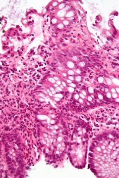
Sarcoidosis
A non-infectious granulomatous disease often affecting lungs, skin, or lymph nodes.
Sarcoidosis, also known as Besnier–Boeck–Schaumann disease, is a non-infectious granulomatous disease characterized by abnormal collections of inflammatory cells that form granulomata. It usually begins in the lungs, skin, or lymph nodes, though any organ can be affected, and often presents with no or only mild symptoms. The condition may resolve without treatment within a few years, but in cases causing significant health problems, steroids such as prednisone are indicated.
Quick Facts
- Field
- Rheumatology, immunology
- Pronounce
- ˌ
- s
- ɑːr
- k
- ɔɪ
- ˈ
- d
- oʊ
- s
- ɪ
- s SAR
- koy
- DOH
- sis
- Onset
- 20–50 years old; More common in women
- Duration
- Few years to long term
- Risks
- Family history
- Diagnosis
- Based on symptoms and tissue biopsy
- Differential
- Tuberculosis
- lymphoma
- infectious mononucleosis
- pulmonary eosinophilia
- Treatment
- Ibuprofen
- prednisone
- methotrexate
Facts from the source article.
Did You Know?
- The heritability of sarcoidosis varies by ethnicity; about 20% of African Americans with the condition have a family member affected, compared to about 5% of European Americans.
- In Swedish individuals, heritability was found to be 39%, and having a first-degree family member with the disease confers a four-fold greater risk.
- Sarcoidosis can develop in people being treated with TNF antagonists like etanercept.
Signs and symptoms
Sarcoidosis is a condition where inflammation can show up anywhere in the body, and roughly 5% of people who have it never notice any symptoms—it gets found by chance. When symptoms do appear, they are often hard to pin down: fatigue that sleep doesn’t fix (affecting up to 85% of patients), low energy, weight loss, joint pain (around 70% of cases), arthritis in 14–38%, dry eyes, swollen knees, blurry vision, shortness of breath, a dry cough, or skin issues. Occasionally, someone might cough up blood. Anxiety, depression, and other psychological distress are common, especially alongside the fatigue. Skin problems can range from rashes and small bumps to erythema nodosum, granuloma annulare, or lupus pernio. The combination of erythema nodosum, swollen lymph nodes in the chest, and joint pain is called Löfgren syndrome, which tends to have a better outlook and is much more common in Scandinavian people. The lungs are involved in at least 90% of cases; about half of those affected end up with lasting lung damage, and 5–15% develop progressive scarring. Doctors use four radiological stages (I through IV) based on the Scadding scale to estimate long-term prognosis, but the scale’s variation makes it unreliable for guiding individual treatment decisions.
Pathophysiology
Granulomatous inflammation in sarcoidosis is characterized primarily by accumulation of macrophages and activated T-lymphocytes, with increased production of key inflammatory mediators such as tumor necrosis factor alpha (TNF), interferon gamma, and several interleukins (IL-2, IL-8, IL-10, IL-12, IL-18, IL-23) and transforming growth factor beta, indicative of a T helper cell-mediated immune response. The disease has paradoxical effects: it features increased macrophage and CD4 helper T-cell activation resulting in accelerated inflammation, yet immune response to antigen challenges such as tuberculin is suppressed, suggesting a state of anergy. Regulatory T-lymphocytes in the periphery of sarcoid granulomas appear to suppress IL-2 secretion, which may cause anergy by preventing antigen-specific memory responses. B cells also likely play a role. Serum levels of soluble human leukocyte antigen class I antigens and angiotensin converting enzyme (ACE) are higher in people with sarcoidosis, and the CD4/CD8 T cell ratio in bronchoalveolar lavage is usually elevated (typically >3.5). Cases have been reported as part of the immune reconstitution syndrome of HIV.
Diagnosis
Diagnosing sarcoidosis involves ruling out other conditions first, as no single test confirms it except the Kveim-Siltzbach test. For lung-related symptoms, doctors may use chest X-rays, CT or PET scans, and various biopsy methods like bronchoscopy, mediastinoscopy, or endoscopic ultrasound with fine-needle aspiration. Biopsied lymph node tissue is checked with flow cytometry to exclude cancer, plus special stains to rule out bacteria and fungi. Blood tests can measure markers such as angiotensin-converting enzyme, which helps track the disease, along with serum amyloid A, soluble interleukin-2 receptor, lysozyme, and KL-6. A bronchoalveolar lavage showing a CD4/CD8 T cell ratio of 3.5 or higher suggests pulmonary sarcoidosis but isn’t definitive. Conditions that can mimic it include lymphoma, metastatic cancer, tuberculosis, fungal infections, rheumatoid nodules, and granulomatosis with polyangiitis. Chest X-ray findings are grouped into four stages: bihilar lymphadenopathy; that plus reticulonodular infiltrates; bilateral pulmonary infiltrates; and fibrocystic disease with upward hilar retraction. However, these stages don’t represent a progression and are mainly of historical interest.
Treatment
Treatments vary greatly; at least half of patients require no systemic therapy, and most (>75%) only need symptomatic treatment with nonsteroidal anti-inflammatory drugs like ibuprofen or aspirin. For active pulmonary sarcoidosis without devastating respiratory impairment, observation without therapy for two to three months is typical; if inflammation does not subside spontaneously, therapy is instituted. Major drug categories include glucocorticoids, antimetabolites, and biologic agents such as monoclonal anti-tumor necrosis factor antibodies. Corticosteroids (most commonly prednisone or prednisolone) have been the standard for many years, though some people do not respond, and their use in mild disease is controversial because many cases remit spontaneously. Antimetabolites (steroid-sparing agents) such as azathioprine, methotrexate, mycophenolic acid, and leflunomide are used as alternatives. Methotrexate is most widely used and studied, considered first-line in neurosarcoidosis often with corticosteroids; long-term use is associated with liver damage in about 10% of people and can cause pulmonary toxicity, so it is often combined with folic acid. Azathioprine can also cause liver damage, but the risk of infection appears about 40% lower with methotrexate.
More in Steroid-responsive Inflammatory Conditions
Spotted an error? Know more?
Reader corrections go straight into our review queue. Suggest an edit · How this site is sourced