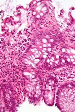
Relapsing polychondritis
A systemic disease with recurrent cartilage inflammation and potential life-threatening complications.
This systemic condition causes repeated flare-ups of cartilage inflammation and sometimes damage. It can turn life-threatening if it affects the windpipe, heart valves, or blood vessels. The exact cause remains unclear.
Quick Facts
- Field
- Rheumatology, Immunology
Facts from the source article.
Did You Know?
- Relapsing polychondritis is an autoimmune disease in which the body's immune system attacks and destroys cartilage tissues; both cell-mediated and humoral immunity are thought to be responsible.
- In 2017, a physician who also had the disease became a rheumatologist to study it, leading to increased awareness and research advances.
- If inflamed cartilage presses against nerves, it can cause problems such as peripheral neuropathy.
Signs and symptoms
Though any cartilage may be affected, the disease often involves several areas while sparing others. Variable signs and symptoms can delay recognition for months, years, or decades. About one-third of people with RP may have associated autoimmune diseases, vasculitides, or hematologic disorders; systemic vasculitis is the most common association, followed by rheumatoid arthritis and systemic lupus erythematosus. Relapsing chondritis is characteristic. Ear cartilage inflammation is a specific symptom, affecting about 20% at presentation and 90% at some point; both ears are often affected, with the entire outer ear except the earlobe becoming swollen, red or purplish, warm, and painful. Inflammation usually lasts days to weeks, resolves spontaneously, and recurs. After several flares, cauliflower ear deformity may develop in about 10% of persons. Nasal cartilage inflammation involves the bridge of the nose, present in 15% at presentation and 65% at some point; nasal obstruction is uncommon. Atrophy may develop gradually, leading to painless but irreversible saddle-nose deformity.
Diagnosis
No specific test exists for relapsing polychondritis. Some people have abnormal lab results, while others have normal labs even during active flares. Patients with ear and nose chondritis and hematologic abnormalities such as macrocytic anemia and thrombocytopenia should be tested for VEXAS syndrome. Clinical criteria from McAdam et al. (1976), expanded by Damiani et al. (1979), and modified by Michet et al. (1986) are used. Acute episodes often show high ESR or CRP. Cartilage-specific antibodies may be present during acute episodes. Antinuclear antibody reflexive panel, rheumatoid factor, and antiphospholipid antibodies can assist evaluation. FDG PET may detect early disease; MRI, CT, and X-rays may reveal inflammation or damaged cartilage. Biopsy of cartilage may show chondrolysis, chondritis, and perichondritis but does not confirm diagnosis. Pulmonary function tests, including flow-volume loops, are usually not useful early but can quantify extrathoracic airway obstruction in later disease.
Treatment
Treatments for relapsing polychondritis lack large-scale trials; doctors rely on smaller studies and individual cases. Mild cases may improve with NSAIDs, while severe ones often need corticosteroids. Dapsone at 25 to 200 mg daily has shown benefit in multiple reports. Drugs like azathioprine or methotrexate can help reduce steroid doses and side effects. For severe illness, cyclophosphamide is frequently combined with high-dose intravenous corticosteroids.
History
In 1923, Rudolf Jaksch von Wartenhorst identified relapsing polychondritis in Prague, first calling it Polychondropathia. His patient, a 32-year-old brewer, had fever, asymmetric polyarthritis, and painful, swollen ears and nose. The name Relapsing Polychondritis, or RP, was given by Pearson and his team in 1960 to highlight its recurring nature.
More in Steroid-responsive Inflammatory Conditions
Spotted an error? Know more?
Reader corrections go straight into our review queue. Suggest an edit · How this site is sourced