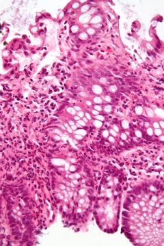
Microscopic colitis
Two related conditions causing chronic watery diarrhea with normal colonoscopy.
Microscopic colitis encompasses two related conditions, collagenous colitis and lymphocytic colitis, that cause chronic non-bloody watery diarrhea. The colon appears normal during colonoscopy, but characteristic inflammatory changes are visible on histopathology. It is notable as a diagnosis in approximately 10% of chronic non-bloody diarrhea cases.
Quick Facts
- Field
- Gastroenterology
- Symptoms
- Persistent diarrhea
- Types
- Lymphocytic colitis, collagenous colitis
- Diagnosis
- Endoscopic biopsy of colonic mucosa
- Differential
- Bile acid diarrhea
- celiac disease
- lactose malabsorption
- Crohn's disease
- ulcerative colitis
- infectious colitis
- Medication
- Loperamide
- Bismuth subsalicylate
- Budesonide
- Frequency
- 103 cases per 100,000 persons
Facts from the source article.
Did You Know?
- Bile acid diarrhea is found in 41% of patients with collagenous colitis and 29% with lymphocytic colitis.
- The two types share many features, including epidemiology, risk factors, and response to therapy, suggesting they are subtypes of the same disease.
- The prognosis is good; most people recover from diarrhea and histological abnormalities resolve, though relapses are common without maintenance treatment.
Signs and symptoms
The hallmark of microscopic colitis is ongoing, watery diarrhea without blood, which can be severe. Other symptoms include belly pain, loss of bowel control, and dropping weight without trying. People with this condition more often have autoimmune diseases like arthritis, Sjögren’s, thyroid issues, or celiac disease. Certain medications—including proton pump inhibitors, H2 blockers, SSRIs, and NSAIDs—are linked to it, and smoking is a major risk factor.
Diagnosis
Colonoscopic appearances are normal or near normal, and a full colonoscopy is required because changes are often patchy. Multiple colonic biopsies are taken; histological features include more than 20 intraepithelial lymphocytes per 100 epithelial cells. In collagenous colitis, a thickened subepithelial collagen band of 10–20 μm is additionally present. Inflammation of the lamina propria with mainly mononuclear cells may be observed. Differential diagnoses to rule out include bile acid diarrhea, lactose malabsorption, celiac disease, Crohn's disease, ulcerative colitis, and infectious colitis.
Treatment
Budesonide, a glucocorticoid formulated for activity in the distal colon and rectum, has been shown in randomized placebo-controlled trials to be effective for both active disease and relapse prevention, though relapse frequently occurs after withdrawal. Other agents such as antidiarrheals, bismuth subsalicylate, mesalazine, systemic corticosteroids, cholestyramine, immunomodulators, and probiotics are less effective. Anti-TNF inhibitors, split ileostomy, diverting ileostomy, and subtotal colectomy are options for steroid-dependent or refractory cases.
Epidemiology
Microscopic colitis is nearly as common as ulcerative colitis and Crohn's disease, with about 103 cases per 100,000 people. It mostly affects middle-aged women, diagnosed on average at age 65, though a quarter of cases occur before age 45.
More in Steroid-responsive Inflammatory Conditions
Spotted an error? Know more?
Reader corrections go straight into our review queue. Suggest an edit · How this site is sourced