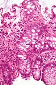
Dermatomyositis
Autoimmune disease affecting skin and muscle with distinct autoantibodies.
Dermatomyositis is a systemic autoimmune inflammatory condition that primarily affects the skin and skeletal muscles, often presenting with a characteristic rash and progressive muscle weakness. It is notable for its association with distinct myositis-specific autoantibodies that define clinical subtypes and directly contribute to disease pathogenesis. The condition can occur as a paraneoplastic syndrome and has a generally favorable prognosis with timely treatment.
Quick Facts
- Field
- Rheumatology
- Symptoms
- Rash
- muscle weakness
- weight loss
- fever
- Complications
- Calcinosis
- dysphagia
- interstitial lung disease
- heart disease (rarely)
- joint pain
- other autoimmune conditions
- Onset
- 40s to 50s
- Duration
- Long term
- Causes
- Autoimmune (Type III hypersensitivity)
- Risks
- Other autoimmune conditions
- ovarian cancer
- breast cancer
- lung cancer
- other cancers
- Diagnosis
- Based on symptoms
- blood tests
- electromyography
- muscle biopsies
Facts from the source article.
Did You Know?
- Opera singer Maria Callas allegedly had dermatomyositis from 1975 until her death.
- Actor Laurence Olivier had dermatomyositis from 1974 until his death.
- American football running back Ricky Bell died at age 29 from heart failure caused by this disease.
Signs and symptoms
The main symptoms include several kinds of skin rash along with muscle weakness in both upper arms or thighs. One form the rashes take is called heliotrope, a purplish or lilac color that may also be red, occurring around the eyes with swelling, as well as on the upper chest or back in a shawl pattern or V-sign above the breasts, and may also appear on the face, upper arms, thighs, or hands. Another form is Gottron's sign, red or violet, sometimes scaly, slightly raised papules on the finger joints or over other bony prominences such as the elbows, knees, or feet. These rashes are worsened by sunlight and are often very itchy, painful, and may bleed. People with only skin findings without weakness or abnormal muscle enzymes may be classified as having amyopathic or clinically amyopathic dermatomyositis. Muscle weakness progressively worsens in proximal muscles like the shoulders and thighs, making tasks such as standing from sitting, lifting, and climbing stairs increasingly difficult. Respiratory failure primarily results from interstitial lung disease, especially in patients with anti-MDA5 autoantibodies who are at high risk of rapidly progressive interstitial lung disease. Respiratory symptoms occur in about 40% of people and may slowly progress, increasing morbidity and mortality. Myocarditis and cardiac conduction system abnormalities can occur but typically have no symptoms or clinical consequences.
Etiopathogenesis
Recent studies indicate that the pathogenesis of dermatomyositis is driven by the pathogenic internalization of autoantibodies, which target intracellular proteins but can enter different cell types and disrupt the function of their target autoantigens, causing inflammation and damage. For example, anti-Mi-2 autoantibodies bind PHD-containing proteins, including a component of the NuRD complex, inducing derepression of multiple genes, while anti-MDA5 autoantibodies directly activate MDA5, inducing type I interferon pathways. The type I interferon pathway is especially prominent and has become a major therapeutic target, supported by clinical responses to JAK inhibitors, anti-IFNβ therapy, and agents targeting the interferon receptor. The characteristic pathological feature is perifascicular muscle involvement, often with vasculopathy, and plasma cells near affected areas externalize immunoglobulin RNA into surrounding muscle cells, suggesting a mechanism for immunoglobulin entry. Dermatomyositis is paraneoplastic in up to 40% of cases, most commonly with anti-TIF1γ autoantibodies, and a majority of such patients harbor somatic mutations in tumor genes encoding the target autoantigens. Inherited genetic factors, including HLA subtypes HLA-DR3, HLA-DR52, and HLA-DR6, predispose to the disease.
Diagnosis and classification
Contemporary diagnostic and classification criteria for dermatomyositis combine clinical features with myositis-specific and myositis-associated autoantibody testing, supported by laboratory, imaging, histopathologic, and electrophysiologic investigations. Principal diagnostic features include detection of myositis-specific autoantibodies such as anti-Mi-2, anti-NXP2, anti-TIF1-γ, anti-MDA5, and anti-SAE, which are considered pathogenic and typically mutually exclusive. Other features include muscle weakness in both thighs or both upper arms; elevated levels of muscle enzymes such as creatine kinase, aldolase, and transaminases; electromyography findings of erratic, repetitive high-frequency signals, short low-energy signals with multiple phases, and sharp activity upon needle insertion; and muscle biopsy showing perifascicular atrophy, increased type I interferon-inducible markers, mononuclear white blood cells between muscle cells, and abnormal degeneration and regeneration. Rashes typical of dermatomyositis, including heliotrope rash, Gottron's sign, and Gottron's papules, are also key. Patients with antisynthetase autoantibodies such as anti-Jo-1 who present with dermatomyositis-like skin manifestations are now generally considered to have antisynthetase syndrome rather than dermatomyositis.
Treatment
Standard treatment for dermatomyositis typically combines glucocorticoids with steroid-sparing immunosuppressive agents such as methotrexate, mycophenolate mofetil, azathioprine, tacrolimus, and cyclosporine. Intravenous immunoglobulin has demonstrated efficacy, and rituximab remains an important option for refractory disease despite mixed clinical trial results. Janus kinase inhibitors including tofacitinib, ruxolitinib, baricitinib, and brepocitinib have shown efficacy, and brepocitinib was approved for medical use in the United States in August 2026. Additional strategies targeting the type I interferon pathway, such as blockade of interferon-β with dazukibart, have also shown benefit. Emerging therapies include CD19 chimeric antigen receptor T-cell therapy, plasma cell-directed therapies, and neonatal Fc receptor inhibitors such as efgartigimod. Antimalarial medications like hydroxychloroquine are generally limited to patients who have previously responded well and tolerated them.
Frequently Asked Questions
What causes Dermatomyositis?
Listed causes of Dermatomyositis include autoimmune (Type III hypersensitivity).
How is Dermatomyositis diagnosed?
Diagnosis of Dermatomyositis is based on based on symptoms, blood tests, electromyography and muscle biopsies.
More in Steroid-responsive Inflammatory Conditions
Spotted an error? Know more?
Reader corrections go straight into our review queue. Suggest an edit · How this site is sourced