
Dermatomyositis
Systemic autoimmune disease affecting skin, muscles, and often internal organs.

Elizabeth M Dugan, Adam M Huber, Frederick W Miller, Lisa G Rider · CC BY-SA 3.0
Dermatomyositis is a group of systemic autoimmune conditions that mainly target the skin and skeletal muscles. The hallmark signs are a distinctive skin rash and muscle weakness that gets worse over time. These symptoms can appear suddenly or develop gradually over months. Other possible symptoms include weight loss, fever, lung inflammation, and sensitivity to light. Complications can involve calcium deposits forming in the skin or muscles. The disease is driven by specific autoantibodies called myositis-specific autoantibodies (MSAs), which define different subtypes of dermatomyositis. Each subtype has its own set of symptoms, outlook, and response to treatment. About 50–80% of adults and 60–70% of children with the juvenile form have one of these MSAs. These autoantibodies are made by plasma cells that have infiltrated the tissues; they are primarily considered markers of disease subsets, and their direct role in entering cells and causing damage is not established. Dermatomyositis can also appear as a paraneoplastic syndrome linked to certain cancers. In these cases, the tumors often have genetic changes, including somatic mutations, in the genes that code for the autoantigens targeted by the patient's own antibodies. Several viruses, especially coxsackievirus, have been associated with the disease, but no definite cause-and-effect link has been proven. Diagnosis usually involves a combination of symptoms, blood tests, electromyography, and muscle biopsies.
Current treatment relies on immunosuppressive drugs like corticosteroids, methotrexate, mycophenolate mofetil, azathioprine, tacrolimus, and cyclosporine. Intravenous immunoglobulin (IVIG) is often used for severe or hard-to-treat cases. Rituximab remains an option, even though clinical trial results have been mixed. Newer targeted therapies, such as Janus kinase (JAK) inhibitors and drugs aimed at the type I interferon pathway (like dazukibart), have shown promising results. Experimental approaches—including CD19 chimeric antigen receptor (CAR) T-cell therapy, treatments that target plasma cells, and neonatal Fc receptor (FcRn) inhibitors like efgartigimod—are also showing encouraging signs. In particular, CD19 CAR-T therapy may offer the possibility of lasting disease control without ongoing treatment. With early diagnosis and proper care, the outlook for dermatomyositis is generally good.
- First described
- 1800s
- Annual incidence
- about 1 in 100,000 people
- Typical age of onset
- 40s and 50s
- Sex predominance
- women more often than men
- Key autoantibodies
- myositis-specific autoantibodies (MSAs)
- Common associated cancers
- ovarian, breast, lung
Lore & Background
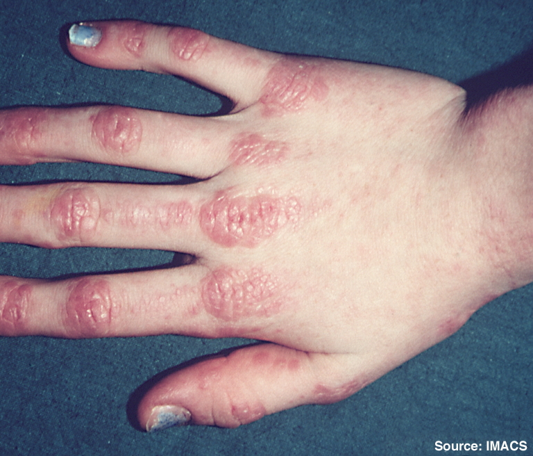
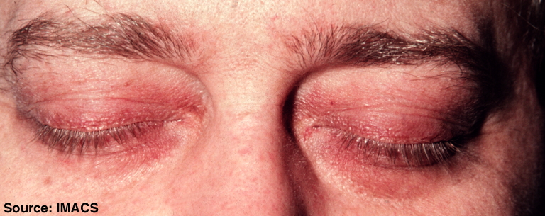
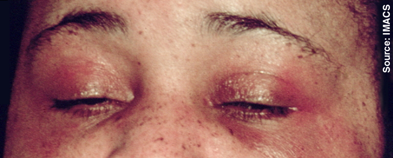
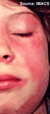
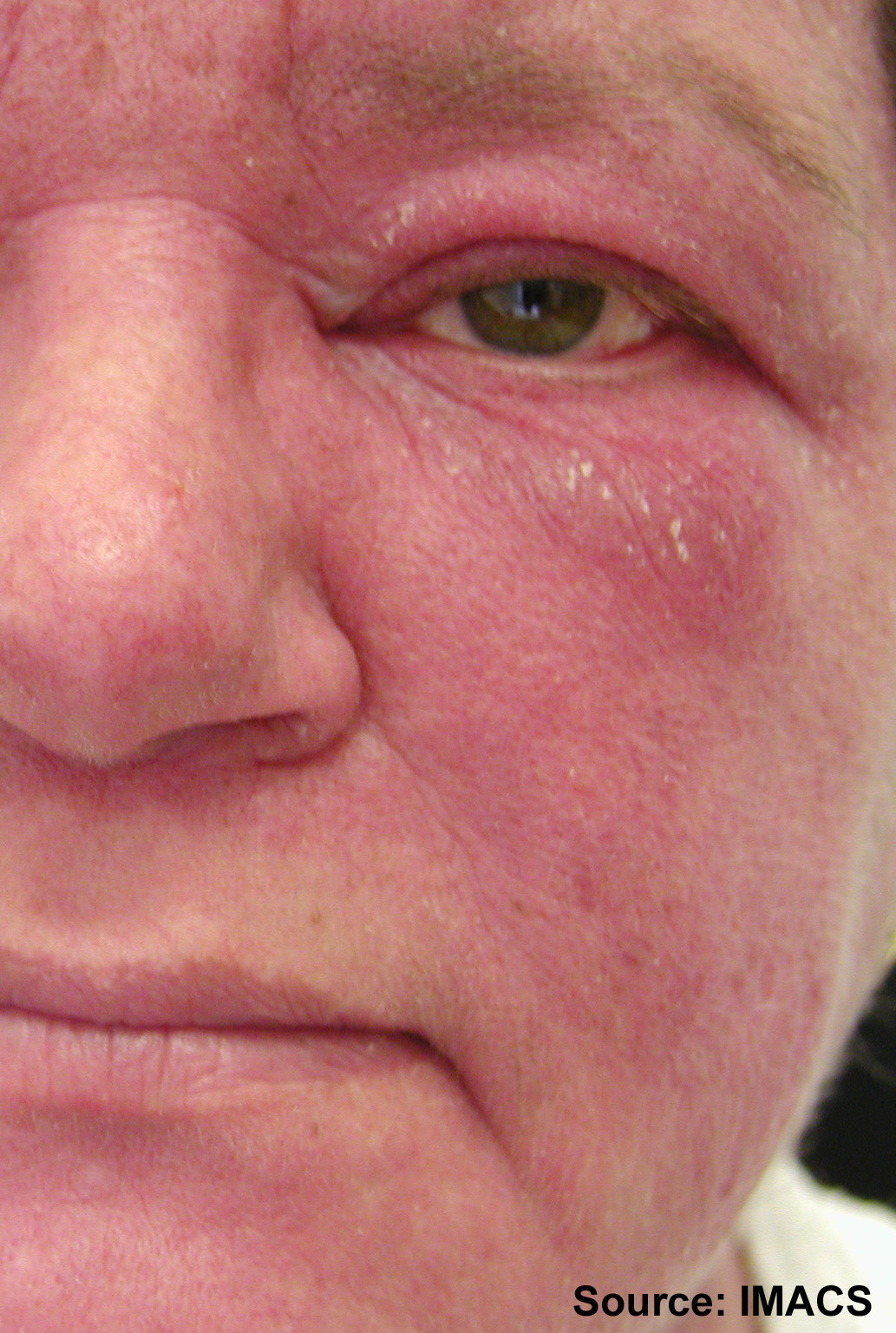
Dermatomyositis presents with characteristic skin rashes, including heliotrope (purplish) discoloration around the eyes and Gottron's papules over finger joints. Muscle weakness typically affects the proximal muscles of the shoulders and thighs, making tasks like standing from sitting or climbing stairs difficult. About 40% of patients develop respiratory symptoms, primarily interstitial lung disease, which is especially prominent in those with anti-MDA5 autoantibodies.
Reader's Guide
Dermatomyositis is significant as a prototypical autoimmune disease where myositis-specific autoantibodies directly drive pathogenesis by entering cells and disrupting target autoantigens. Its association with malignancies in up to 40% of cases, particularly in patients with anti-TIF1γ autoantibodies, highlights its role as a paraneoplastic syndrome. The condition's prognosis is generally favorable with timely diagnosis and appropriate treatment, which includes immunosuppressive agents and emerging targeted therapies such as JAK inhibitors and CD19 CAR-T cell therapy. The discovery that tumors in paraneoplastic cases harbor somatic mutations in genes encoding the targeted autoantigens suggests an anti-tumor immune response gone awry.
The Visible and Invisible Hallmarks
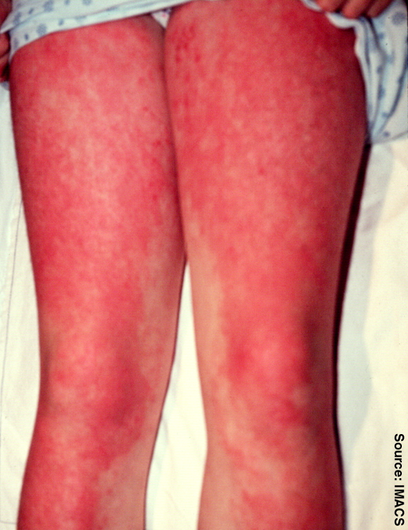
Dermatomyositis announces itself through a constellation of skin and muscle findings that can appear suddenly or creep in over months. The most recognizable cutaneous markers include a purplish or lilac discoloration around the eyes—known as the heliotrope rash—along with a reddened shawl pattern across the upper back and neck or a V-sign above the breasts. Gottron's papules, small raised red or violet bumps, cluster over finger joints, elbows, knees, and feet. All of these rashes worsen under sunlight and can become intensely itchy, painful, or even bleed. Muscularly, patients face a progressive weakening of proximal muscles in the shoulders and thighs, making everyday tasks like rising from a chair or climbing stairs increasingly arduous. Beyond the skin and limbs, roughly forty percent develop respiratory compromise from interstitial lung disease, about thirty percent experience mild joint swelling, and many later struggle with swallowing or reflux. Diagnosis typically weaves together clinical observation, blood work, electromyography, and muscle biopsy. A variant called amyopathic dermatomyositis presents with characteristic rashes but no measurable muscle weakness or enzyme elevation.
The Autoantibody Engine and Disease Subtypes
At the molecular core of dermatomyositis lies a self-directed immune response in which plasma cells produce myositis-specific autoantibodies that target intracellular proteins. Remarkably, these antibodies do not merely circulate; they infiltrate various cell types and disrupt the function of their target autoantigens, directly triggering cellular damage and inflammation. This pathogenic internalization drives the disease process. Approximately eighty percent of adult patients and sixty percent of children with juvenile dermatomyositis carry at least one such antibody, and each distinct profile defines a clinically and pathologically separate subtype with its own expected course, risk profile, and treatment response. For instance, anti-Mi-2 antibodies bind PHD-containing proteins within the NuRD complex and induce derepression of multiple genes, while anti-MDA5 antibodies are strongly linked to rapidly progressive interstitial lung disease. Dermatomyositis can also emerge as a paraneoplastic syndrome, where tumors harbor somatic mutations in the very genes encoding the autoantigens the patient's antibodies attack. Although coxsackievirus and other infections have been associated with the condition, no definitive causal link has been established.
A Shifting Therapeutic Landscape
Managing dermatomyositis currently depends on a tiered immunosuppressive strategy. First-line agents include corticosteroids alongside steroid-sparing drugs such as methotrexate, mycophenolate mofetil, azathioprine, tacrolimus, and cyclosporine. For patients whose disease proves refractory or particularly severe, intravenous immunoglobulin has become a widely employed rescue therapy. Rituximab occupies an important but somewhat ambiguous niche, as clinical trial results have been mixed. The frontier of treatment is expanding rapidly: Janus kinase inhibitors and agents that block the type I interferon pathway, exemplified by dazukibart, have shown promising efficacy in early studies. Even more novel approaches are entering the picture—CD19 chimeric antigen receptor T-cell therapy, plasma cell–directed strategies, and neonatal Fc receptor inhibitors like efgartigimod are all demonstrating encouraging results. Perhaps most strikingly, CD19 CAR-T cell therapy offers the tantalizing possibility of sustained, treatment-free disease control, a concept that would represent a paradigm shift in how chronic autoimmune conditions are managed.
Who Is Affected and What the Future Holds
Dermatomyositis is a rare condition, with roughly one in every one hundred thousand people receiving a new diagnosis each year. It most commonly strikes individuals in their fourth and fifth decades of life, and women are affected more frequently than men, though people of any age can develop the disease. The condition was first described in the nineteenth century, and its clinical recognition has evolved considerably since then. Despite the seriousness of its potential complications—including calcium deposits in muscle or skin, life-threatening interstitial lung disease, and occasionally severe cardiac involvement such as arrhythmias or heart failure—the overall prognosis is generally favorable when the disease is identified promptly and treated appropriately. The availability of distinct autoantibody subtypes now allows clinicians to anticipate which patients are at higher risk for particular complications, such as the rapidly progressive lung disease seen with anti-MDA5 positivity. This precision in risk stratification, combined with the expanding arsenal of targeted therapies, suggests that the outlook for newly diagnosed patients continues to improve with each passing year.
Gallery
Frequently Asked Questions
Who is Dermatomyositis?
Dermatomyositis is a systemic autoimmune condition that primarily attacks the skin and skeletal muscles, with its first clinical descriptions dating back to the 1800s. It is not a single disease but a group of related conditions unified by their shared target organs.
What are Dermatomyositis's powers/role?
Its signature moves include a distinctive skin rash and progressively worsening muscle weakness, which can emerge suddenly or creep in over several months. Additional manifestations may include fever, unexplained weight loss, lung inflammation, and heightened sensitivity to sunlight.
Why is Dermatomyositis important?
It is driven by a specific set of myositis-specific autoantibodies (MSAs) that carve the condition into distinct subtypes, making those markers central to diagnosis and classification. With an annual incidence of roughly one in 100,000 people, it remains a rare but clinically significant entity in rheumatology.
Who does Dermatomyositis usually target?
The condition shows a clear sex bias, affecting women more frequently than men, and typically declares itself during the fourth or fifth decade of life. Its onset in the 40s and 50s makes it a notable concern for middle-aged adults seeking a rheumatology workup.
More in Steroid-responsive inflammatory conditions 1-24
Spotted an error? Know more?
Reader corrections go straight into our review queue. Suggest an edit · How this site is sourced