
Cerebral vasculitis
Inflammation of brain blood vessels causing stroke and varied symptoms.

Marvin 101 · CC BY-SA 3.0
Cerebral vasculitis, also termed angiitis, is a condition characterized by inflammation of the blood vessel walls within the brain and occasionally the spinal cord. It can affect capillaries, arterioles, venules, arteries, and veins, and may lead to stroke if blood flow is reduced or stopped. The disorder produces a wide range of neurological symptoms, including headache, cognitive impairment, focal deficits, and seizures, and can mimic multiple sclerosis.
- Incidence
- 2.4 cases per 1 million people
- Mortality
- 8–23%
- Severe disability risk
- 25%
- Common presenting symptoms
- focal neurological deficits (63%), headaches (51%), cognitive impairment (41%)
- Associated bleeding
- 10% have brain bleeding
- Treatment response
- 75% have favorable response to therapy
- Long term remission
- 21–66%
Lore & Background
Cerebral vasculitis is classified as primary when no secondary cause is identified, with the exact mechanism unknown. Secondary causes include infections such as varicella zoster virus, systemic autoimmune diseases like lupus and rheumatoid arthritis, medications and drugs (amphetamine, cocaine, heroin), certain cancers (lymphomas, leukemia, lung cancer), and other systemic vasculitides. The condition can imitate and be imitated by diseases such as fibromuscular dysplasia and thrombotic thrombocytopenic purpura.
Diagnosis relies on brain imaging and biopsy, with digital subtraction angiography being the preferred imaging modality for medium to large vessel disease. Biopsy is the most definitive test but has a low yield (30–50% normal in suspected cases) due to irregular vessel involvement or risk of bleeding. High-resolution MRI may show vessel wall hyperenhancement and multiple infarcts, and a normal MRI makes the diagnosis unlikely. Inflammatory markers are usually normal, and cerebrospinal fluid analysis shows nonspecific mild leukocytosis and increased protein.
Treatment primarily involves glucocorticoids and cyclophosphamide, with induction followed by at least two years of maintenance therapy. Aspirin may be added for medium or large vessel involvement. Prognosis varies: 21–66% achieve long-term remission, 12–59% have flares, and 46–73% retain good neurological function. Poor prognosis factors include older age, cognitive impairment at diagnosis, cerebral infarctions, spinal cord involvement, diagnostic delays, and medium/large vessel involvement.
Reader's Guide
Cerebral vasculitis is a rare but serious inflammatory disorder of the central nervous system blood vessels, with an incidence of 2.4 per million. Its significance lies in its potential to cause stroke, cognitive decline, and severe disability, with a mortality of 8–23% and a 25% risk of severe disability. The condition's varied presentation—mimicking multiple sclerosis, encephalopathy, or mass lesions—makes diagnosis challenging, often requiring angiography and biopsy. Treatment with immunosuppressants yields favorable responses in 75% of cases, but long-term remission rates vary widely (21–66%). The disorder underscores the importance of recognizing secondary causes, such as infections, autoimmune diseases, and drugs, and highlights the need for prompt diagnosis to improve outcomes. Its legacy includes advancing understanding of CNS inflammation and the development of targeted therapies like rituximab and tocilizumab for refractory cases.
Did You Know?
- Cerebral vasculitis can affect all blood vessel sizes, from capillaries to large arteries and veins.
- 10% of cases have associated bleeding in the brain.
- Primary CNS vasculitis has an incidence of 2.4 cases per 1 million people.
- Digital subtraction angiography is positive in only 33% of biopsy-confirmed cases.
Clinical Presentation & Symptom Spectrum
Cerebral vasculitis presents with an extraordinarily broad neurological picture, making early recognition difficult. The most commonly reported initial manifestations in primary central nervous system vasculitis include focal neurological deficits observed in roughly 63 percent of patients, persistent headaches in about 51 percent, and measurable cognitive decline in 41 percent. Speech difficulties such as aphasia appear in 35 to 43 percent of cases, while seizures account for 16 to 33 percent. Additional features range from ataxia and visual field loss to acute or subacute encephalopathy that can deteriorate into coma. Because the inflammation can target vessels of every caliber, from capillaries to large arteries and veins, symptoms may include skin rashes, profound fatigue, joint aches, impaired coordination, altered sensation, and even changes in perception, thought, or behavior. In roughly 10 percent of cases the process triggers intracranial bleeding. Some presentations closely mimic multiple sclerosis, and in severe instances a mass-like lesion can develop, precipitating herniation and coma. Spinal cord involvement occurs but remains rare, particularly when it is not accompanied by brain involvement.
Etiology & Prognostic Landscape
Cerebral vasculitis is classified as primary when no identifiable secondary trigger exists, a condition sometimes called primary angiitis of the central nervous system. The precise mechanism behind this primary form remains unknown. Secondary causes, however, span a wide spectrum: viral infections such as varicella zoster, systemic autoimmune conditions including lupus and rheumatoid arthritis, recreational and illicit drugs like amphetamine, cocaine, and heroin, certain malignancies such as lymphomas, leukemia, and lung cancer, and other systemic vasculitides including granulomatosis with polyangiitis, polyarteritis nodosa, and Behçet's disease. The condition can also masquerade as, or be mistaken for, diffuse vascular disorders like fibromuscular dysplasia and thrombotic thrombocytopenic purpura. Epidemiologically, primary central nervous system vasculitis strikes approximately 2.4 individuals per million, carries an 8 to 23 percent mortality rate, and leaves 25 percent of survivors with severe disability. Prognosis worsens significantly with older age, cognitive impairment at diagnosis, visible cerebral infarctions on imaging, spinal cord involvement, diagnostic delays, and inflammation concentrated in medium or large vessels.
Pathological Subtypes & Vessel Involvement
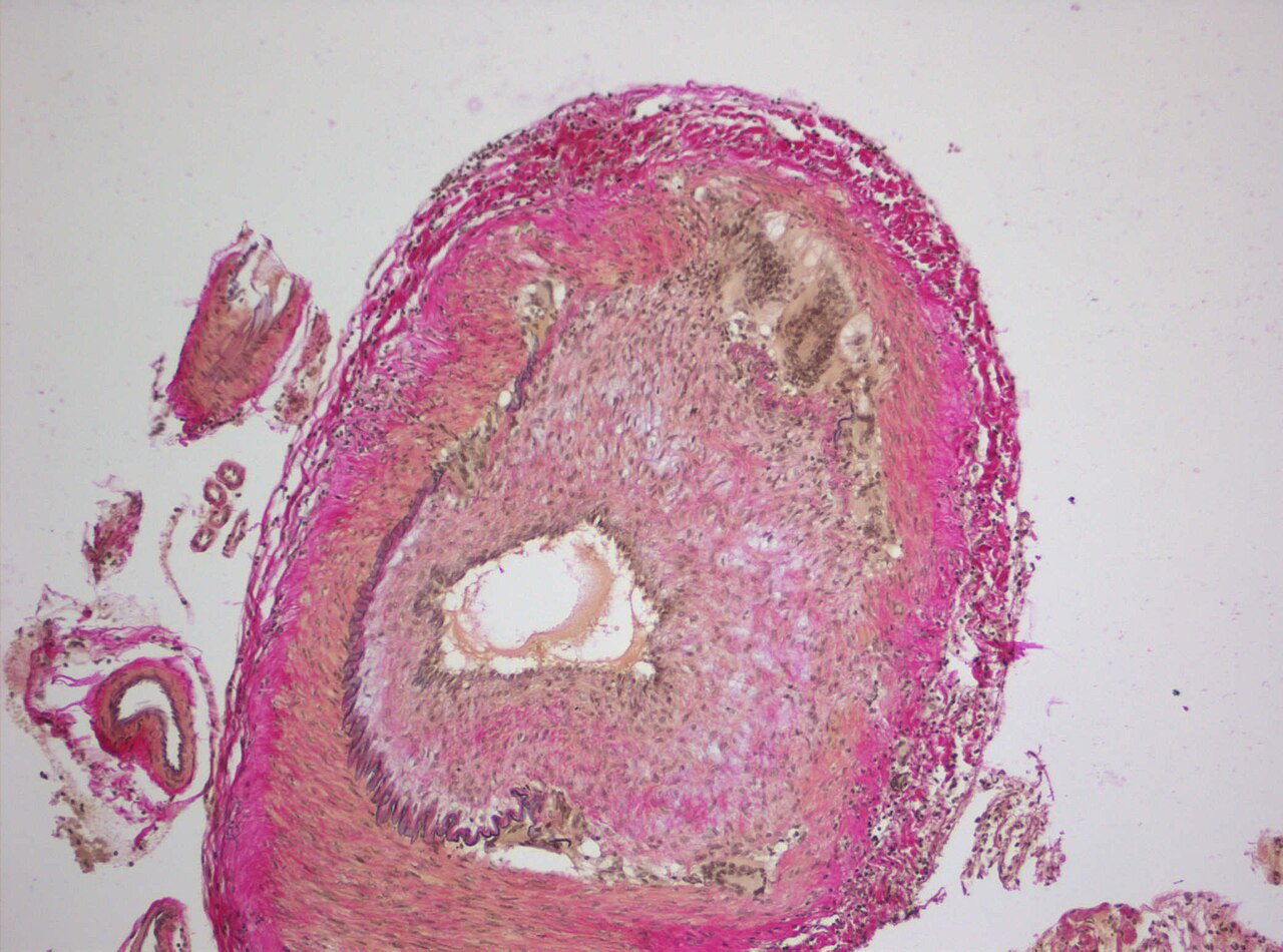
Histological examination of affected vessels in primary central nervous system vasculitis reveals three principal patterns of inflammation. The first, granulomatous vasculitis, identified in 32 to 61 percent of biopsied cases, is characterized by well-organized granulomas containing multi-nucleated giant cells embedded within the vessel wall. The second pattern, lymphocytic vasculitis, found in 24 to 79 percent of cases, features a dense infiltration of lymphocytes into the vessel wall without granuloma formation; notably, this subtype carries a comparatively lower risk of disability and death. The third, necrotizing vasculitis, present in 14 to 42 percent of cases, involves full-thickness transmural fibrinoid necrosis of the vessel wall and is strongly linked to a high probability of intracerebral hemorrhage. The disease does not discriminate by vessel size: capillaries, arterioles, venules, arteries, and veins can all be affected. When the inflammatory process narrows or occludes a vessel, downstream tissue is deprived of oxygen and nutrients, leading to cell death and, in the brain, a stroke.
Diagnostic Complexity & Investigative Limitations
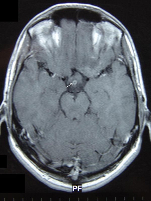
Confirming a diagnosis of primary central nervous system vasculitis remains one of neurology's most challenging endeavors. Although vessel biopsy is considered the gold standard, up to 30 to 50 percent of biopsies in suspected cases return normal, owing to the patchy, irregular distribution of inflamed segments and the practical impossibility of safely sampling larger vessels without risking hemorrhage. Digital subtraction angiography is the preferred imaging tool, offering greater specificity than magnetic resonance or CT angiography for detecting luminal irregularities in medium-sized vessels and the posterior circulation. Typical angiographic findings include smooth, segmental narrowing of multiple arteries, sometimes with post-stenotic dilation or bleeding. However, in small-vessel disease angiography is frequently unremarkable, necessitating biopsy. Crucially, the two modalities show poor concordance: one meta-analysis found angiography positive in only 33 percent of biopsy-confirmed cases, while biopsy was positive in merely 8 percent of angiographically confirmed cases. High-resolution MRI can reveal vessel wall hyperenhancement, multiple infarcts, leptomeningeal enhancement, and intracranial hemorrhages, and a normal MRI effectively argues against the diagnosis. Inflammatory blood markers such as CRP and ESR are typically unremarkable, and cerebrospinal fluid analysis yields only a nonspecific picture of mildly elevated white cells and protein.
Gallery
Frequently Asked Questions
Who is Cerebral vasculitis?
Cerebral vasculitis (also called angiitis) is a rare inflammatory disorder that targets the walls of blood vessels inside the brain and, at times, the spinal cord. It can strike capillaries, arterioles, venules, arteries, and veins, and is diagnosed in roughly 2.4 out of every million people.
What are Cerebral vasculitis's powers/role?
Its 'powers' show up as a broad neurological symptom set: focal deficits appear in about 63% of cases, headaches in 51%, and cognitive impairment in 41%, with seizures also in the mix. In the most dangerous storyline, reduced or halted blood flow triggers a stroke, and roughly 10% of patients additionally suffer brain hemorrhage.
Why is Cerebral vasculitis important?
It is a critical diagnostic puzzle because its symptom profile—headache, seizures, cognitive changes, focal deficits—can closely mimic multiple sclerosis, tripping up even experienced clinicians. Catching it early is essential, since untreated vessel inflammation can escalate to stroke or fatal bleeding.
What's Cerebral vasculitis's weakness?
Its greatest vulnerability is corticosteroid and immunosuppressive therapy, which produces a favorable response in roughly three-quarters of patients. That steroid-responsiveness is exactly what earns it a spot in the 'steroid-responsive inflammatory' category.
More in Steroid-responsive inflammatory conditions 1-24
Spotted an error? Know more?
Reader corrections go straight into our review queue. Suggest an edit · How this site is sourced