
Skin grafting
Surgical transplantation of skin to repair damaged tissue.

Jmelendres · CC BY 3.0
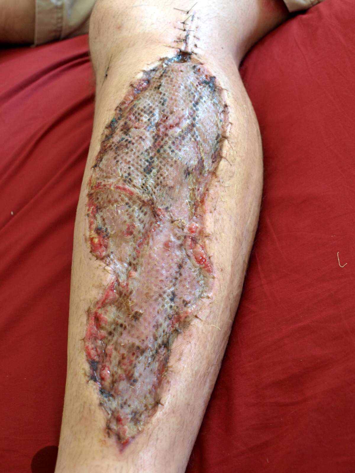
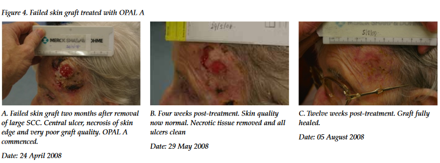
Skin grafting is a surgical procedure where skin is transplanted from one area of the body to another, without its own blood supply. The moved piece of skin is called a skin graft. This technique is used to treat serious wounds, burns, large areas of skin lost to infections like necrotizing fasciitis or purpura fulminans, and after certain surgeries—most often the removal of skin cancers—where healing requires new skin.
The process usually follows a serious injury: damaged skin is surgically removed (through excision or debridement), and then a graft is placed. Grafting serves two main purposes: it shortens treatment time and hospital stays, and it improves both the function and appearance of the injured area.
There are two main types of skin grafts. A partial-thickness graft removes a thin layer of skin from a healthy donor site and is more common. A full-thickness graft removes the entire depth of skin down to the fat, and the full piece is placed on the recipient site. Full-thickness grafts carry a higher risk of rejection, but they leave only a scar line on the donor area, similar to a C-section scar. The donor site from a full-thickness graft heals faster and is less painful than a partial-thickness donor site, which must heal by re-epithelialization—a slow, often painful process.
In medical use, two layers of skin made from animal sources have been found helpful for venous leg ulcers.
Grafts are classified by thickness, source, and purpose. By source: autologous (from the same person, also called an autograft), isogeneic (from a genetically identical donor, like an identical twin), allogeneic (from the same species, such as human to human), xenogeneic (from a different species, such as pig skin), and prosthetic (using synthetic materials like metal or plastic). Allografts, xenografts, and prosthetic grafts are usually temporary dressings to prevent infection and fluid loss; the body eventually rejects them, so they must be removed. Autologous grafts and some treated allografts can stay permanently without rejection. Genetically modified pigs can produce skin equivalent to allografts, and tilapia skin is used as an experimental cheap xenograft where pig skin is unavailable, including in veterinary medicine.
By thickness: split-thickness grafts (STSG) include the epidermis and part of the dermis. Their thickness varies by donor site and patient need. They can be run through a skin mesher to create holes, allowing expansion up to four times their size; for larger expansions, the modified MEEK technique allows up to nine times expansion. Split-thickness grafts are common because they cover large areas with a low rejection rate, and the same donor site can be reused after six weeks. The donor site heals by re-epithelialization and needs dressings. Full-thickness grafts include the epidermis and the entire dermis; the donor site is either stitched closed or covered with a split-thickness graft. Composite grafts are small grafts containing skin and underlying cartilage or other tissue, often taken from the ear to reconstruct nasal alar rim defects.
For donor selection, grafts from other animals (heterografts or xenografts) are temporary biologic dressings that the body rejects within days to weeks. They help reduce bacteria and fluid loss in open wounds. For larger tissue loss, a full-thickness graft is used, especially on the face and hand, where minimizing contraction is important. The thicker the graft, the less contraction and deformity.
Cell cultured epithelial autograft (CEA) procedures take skin cells from the patient and grow them into sheets in a lab. Since the cells are the patient’s own, the immune system won’t reject them. But these sheets are very thin—only a few cell layers—so they don’t handle trauma well, and the “take” rate is often less than 100%. Newer methods combine CEA with a dermal matrix for more support, and research is working on combining both in one product. Experimental procedures for burn victims use stem cells in a solution applied with a skin cell gun, with recent success in applying cells without damage.
To remove thin, well-preserved skin strips from the donor, surgeons use a special instrument called a dermatome. This usually produces a split-thickness graft containing the epidermis and only part of the dermis. The dermis left behind at the donor site contains hair follicles and sebaceous glands, which have epidermal cells that gradually grow back to heal the area.
- field
- Surgery
- known_for
- Transplantation of skin to treat wounds, burns, and skin loss
- types
- Partial-thickness and full-thickness grafts
- sources
- Autologous, isogeneic, allogeneic, xenogeneic, prosthetic
Lore & Background
Skin grafting often takes place after serious injuries when some of the body's skin is damaged. Surgical removal (excision or debridement) of the damaged skin is followed by skin grafting. The grafting serves two purposes: reducing the course of treatment needed (and time in the hospital), and improving the function and appearance of the area of the body which receives the skin graft. There are two types of skin grafts: partial-thickness, which involves removing a thin layer of skin from a healthy part of the body (the donor section), and full-thickness, which involves excising a defined area of skin down to the fat. A full-thickness skin graft is more risky in terms of acceptance but leaves only a scar line on the donor section, similar to a Cesarean-section scar.
Reader's Guide
Skin grafting is a significant surgical technique for repairing extensive skin loss from trauma, burns, or infection. It reduces treatment duration and hospital stay while improving function and appearance. Grafts are classified by thickness (split-thickness, full-thickness, composite) and source (autologous, isogeneic, allogeneic, xenogeneic, prosthetic). Autologous grafts can be left permanently, while allografts, xenografts, and prosthetic grafts are usually temporary. The healing process occurs in three stages: plasmatic imbibition, capillary inosculation, and neovascularization. Negative pressure wound therapy is increasingly used to aid graft healing. Skin grafting remains a cornerstone of reconstructive surgery, with ongoing research into cell cultured epithelial autografts and stem cell applications.
Did You Know?
- A split-thickness skin graft can be processed through a skin mesher to expand up to four times its size, and up to nine times using the modified MEEK technique.
- The healing process for skin grafts occurs in three stages: plasmatic imbibition, capillary inosculation, and neovascularization.
- Genetically modified pigs can produce allograft-equivalent skin material, and tilapia skin is used as an experimental cheap xenograft.
- Cell cultured epithelial autograft (CEA) procedures grow new skin cells in sheets from the patient's own cells, avoiding immune rejection.
Clinical Indications and Therapeutic Goals
Skin grafting is a surgical procedure in which tissue is transplanted without its own blood supply to cover areas where the body's natural skin has been lost or destroyed. Surgeons turn to this technique when patients face extensive trauma, severe burns, or aggressive infections like necrotizing fasciitis and purpura fulminans that strip away large patches of skin. It also plays a critical role after the excision of skin cancers, where the resulting wound must be closed for proper healing. The procedure typically follows a first step of debridement or excision of all damaged tissue. Once the healthy wound bed is prepared, the graft is applied to serve two simultaneous goals: shortening the overall course of treatment and reducing hospital stays, while also restoring both the function and the cosmetic appearance of the affected region. In essence, skin grafting bridges the gap between a devastating tissue loss and the body's ability to resume normal daily life.
Classification and Graft Types
Skin grafts are categorized along several axes: thickness, biological source, and intended purpose. By thickness, the most common is the split-thickness graft, which harvests the epidermis plus a portion of the dermis. A special instrument called a skin mesher punches tiny apertures into the graft, letting surgeons stretch it to cover up to four times its original area; a modified technique known as the MEEK method can expand it to nine times. Full-thickness grafts, by contrast, remove the entire dermis down to the fat layer and are reserved for areas where minimizing contraction matters most, such as the face and hands. Composite grafts go a step further, including skin plus underlying cartilage or other tissue, with the ear being a classic donor site for reconstructing nasal rim defects. By source, grafts range from autologous (same patient), isogeneic (genetically identical twins), allogeneic (same species, different individual), xenogeneic (across species, such as pig skin), to fully synthetic prosthetic implants.
Donor Site Harvesting and Recovery
The choice of donor site carries significant consequences for the patient's recovery. In a split-thickness procedure, a dermatome, a specialized surgical blade, shaves off a thin, uniform slice of skin that retains the epidermis and part of the dermis. The dermis left behind is seeded with hair follicles and sebaceous glands, each housing epidermal cells that slowly migrate outward to regenerate a new skin layer. This re-epithelialization process can be prolonged and notably painful, and the exposed donor area is vulnerable to infection, requiring careful dressing and sometimes subcutaneous or topical anesthetic agents for comfort. A full-thickness harvest, on the other hand, excises a defined block of skin down to the fat. The donor wound is then sutured shut or covered with a split-thickness graft, leaving a linear scar reminiscent of a Cesarean-section mark. Paradoxically, although full-thickness grafts carry a higher risk of the recipient site rejecting the tissue, the donor area often heals faster and with less pain than a split-thickness site. The same split-thickness donor region can be re-harvested after approximately six weeks if additional graft material is needed.
Rejection, Temporary Substitutes, and Emerging Techniques
Not all grafts are meant to stay. Allografts, xenografts, and prosthetic implants function primarily as temporary biological dressings that shield an open wound from infection and fluid loss while the body prepares for a definitive repair. Because the immune system recognizes them as foreign, these materials are eventually rejected within days to a few weeks and must be removed. Autologous grafts, drawn from the patient's own body, avoid this problem entirely and can remain permanently. In resource-limited settings, tilapia skin has emerged as an inexpensive experimental xenograft, and genetically modified pigs are being explored as producers of allograft-equivalent skin. On the frontier of regenerative medicine, cell-cultured epithelial autografts grow the patient's own skin cells into thin sheets in a lab, eliminating rejection risk but producing tissue too fragile to withstand trauma, with take rates often below one hundred percent. Researchers are now pairing these cultured sheets with a dermal matrix for added structural support, and experimental skin-cell guns are being tested to spray stem-cell solutions directly onto burn wounds without damaging the cells.
Gallery
Frequently Asked Questions
What is Skin grafting?
Skin grafting is a surgical procedure in which a piece of skin is transplanted to a damaged area of the body. Unlike other grafts, the transplanted skin does not arrive with its own blood supply and must reattach to the recipient site to survive.
What is Skin grafting used to treat?
Surgeons employ skin grafting to close wounds that are too large or complex to heal on their own, such as severe burns, traumatic injuries, and areas of skin lost to conditions like necrotizing fasciitis. It is also a standard step after excising skin cancers to restore the surface.
What types of Skin grafting exist?
The two main categories are partial-thickness grafts, which include the epidermis and part of the dermis, and full-thickness grafts, which carry the entire dermal layer. The choice depends on how much tissue is missing and where the graft is placed.
Where does the donor skin for Skin grafting come from?
Graft tissue can be autologous (from the patient's own body), isogeneic (from a genetically identical twin), allogeneic (from a human donor), xenogeneic (from another species), or prosthetic (synthetic material). Autologous grafts remain the most common in routine practice.
Why is Skin grafting considered important in surgery?
It provides a reliable way to restore a protective skin barrier when the body cannot regenerate tissue on its own. Without this technique, patients with large burns, infections, or post-surgical defects would face prolonged healing, elevated infection risk, and significant functional loss.
More in Medical Procedures And Treatments 1-23
Spotted an error? Know more?
This is a living reference — every entry is fact-audited, and reader corrections feed straight into our audit queue. Suggest an edit · See this site's audit record