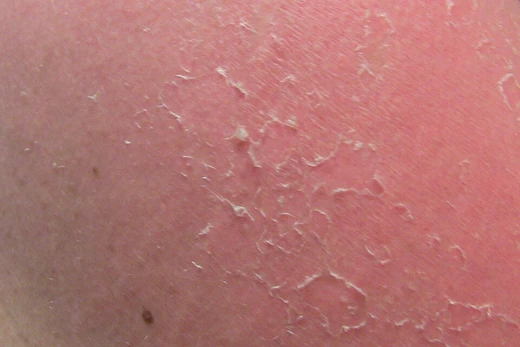
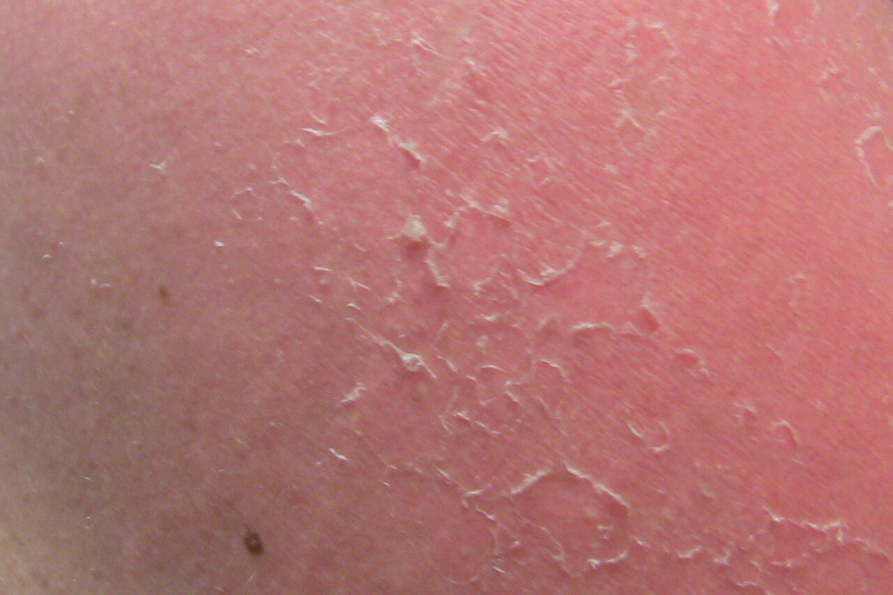
Desquamation
The shedding of dead skin cells from the outermost layer.
Last updated

Barbariandeagle · CC0
The process typically takes about 14 weeks from formation to shedding.[1] The process of dead cells being shed from the skin's outermost layer is referred to as desquamation or peeling skin.[2] When no disease is present, corneocytes are shed one by one from the skin surface without being noticed.[1]
Quick Facts
- Field
- Dermatology
Facts from the source article.
Key facts and history
In pathologic desquamation, such as in X-linked ichthyosis, corneocytes are shed in clusters forming visible scales.[1] Within the epidermis, which is the skin's outermost layer, keratinocytes are the most abundant cell type.[1] The stratum corneum, the epidermis's most superficial layer, consists of corneocytes, which are keratinocytes that have undergone terminal differentiation.[1] Corneodesmosomes are the structures that keep corneocytes attached to one another.[3]
The breakdown of corneodesmosome connections is a necessary step for desquamation to happen.[3] Members of the kallikrein family, particularly KLK1, KLK5, and KLK7, which degrade corneodesmosomes, are produced by keratinocytes in the stratum granulosum.[3] To stop desquamation from occurring too early, keratinocytes in the granular layer also generate proteins that inhibit kallikreins.[3] On acral skin, the desquamation process is slower because granular layer keratinocytes reduce the expression of KLK1 and KLK7.[4]
When desquamation is abnormal in various disease conditions, scale develops on the skin's surface.[1]
What the cited sources add
A 2022 PLOS Computational Biology paper by Miller models desquamation in the interfollicular epidermis.[2] A 1993 paper by Jackson in The Western Journal of Medicine describes the pathobiology of the stratum corneum.[1][3] A 2022 JCI Insight paper by Merleev details biogeographic and disease-specific alterations in epidermal lipid composition and acral keratinocyte analysis.[3][4][5] A 2000 Clinical Microbiology Reviews paper by Dinges discusses Staphylococcus aureus exotoxins in relation to desquamation.[6]
A 2010 eMedicine entry by Parillo covers Stevens-Johnson syndrome.[5] A 2005 CDC publication addresses cutaneous radiation injury.[5]
References
- Wikipedia, "Desquamation" (CC BY-SA 4.0), which cites: Jackson, "Pathobiology of the Stratum Corneum", The Western Journal of Medicine, 1993, p. 279–85 (original not read by us) en.wikipedia.org
- Wikipedia, "Desquamation" (CC BY-SA 4.0), which cites: Miller, "Multiscale modelling of desquamation in the interfollicular epidermis", PLOS Computational Biology, August 2022 (original not read by us) en.wikipedia.org
- Wikipedia, "Desquamation" (CC BY-SA 4.0), which cites: Jackson, "Pathobiology of the Stratum Corneum", The Western Journal of Medicine, 1993, p. 279–85; Merleev, "Biogeographic and disease-specific alterations in epidermal lipid composition and single-cell analysis of acral keratinocytes", JCI Insight, 2022-08-22; Jackson, "Pathobiology of the Stratum Corneum", The Western Journal of Medicine, 1993, p. 279–85; Merleev, "Biogeographic and disease-specific alterations in epidermal lipid composition and single-cell analysis of acral keratinocytes", JCI Insight, 2022-08-22 (original not read by us en.wikipedia.org
- Wikipedia, "Desquamation" (CC BY-SA 4.0), which cites: Merleev, "Biogeographic and disease-specific alterations in epidermal lipid composition and single-cell analysis of acral keratinocytes", JCI Insight, 2022-08-22; Merleev, "Biogeographic and disease-specific alterations in epidermal lipid composition and single-cell analysis of acral keratinocytes", JCI Insight, 2022-08-22 (original not read by us) en.wikipedia.org
- Wikipedia, "Desquamation" (CC BY-SA 4.0), which cites: Parillo, "Stevens-Johnson Syndrome", eMedicine, 2010-05-25; Centers for Disease Control and Prevention, "Cutaneous Radiation Injury", CDC, 2005-06-30; Merleev, "Biogeographic and disease-specific alterations in epidermal lipid composition and single-cell analysis of acral keratinocytes", JCI Insight, 2022-08-22; Merleev, "Biogeographic and disease-specific alterations in epidermal lipid composition and single-cell analysis of acral keratinocytes", JCI Insight, 2022-08-22; Merleev, "Biogeographic and disease-specific alterations in epiderma en.wikipedia.org
- Wikipedia, "Desquamation" (CC BY-SA 4.0), which cites: Dinges, "Exotoxins of Staphylococcus aureus.", Clinical Microbiology Reviews, January 2000, p. 16–34, table of contents (original not read by us) en.wikipedia.org
Each numbered sentence above was checked against the source it cites before publication. Sentences citing Wikipedia adapt text from the English Wikipedia article Desquamation (contributors), used under CC BY-SA 4.0; this entry is available under the same licence. Other sources were used for facts only; no text or images are copied from them.
More in Symptoms and signs: Skin and subcutaneous tissue
Sources
Compiled from Wikipedia and the sources listed below. Text from Wikipedia is available under CC BY-SA 4.0; this entry is adapted from it.
- Wikipedia: Desquamation (CC BY-SA 4.0).
Spotted an error? Know more?
Reader corrections go straight into our review queue. Suggest an edit · How this site is sourced