Cyanosis
A bluish discoloration of tissue indicating low oxygen.

Pots Syndrome · CC BY 2.0
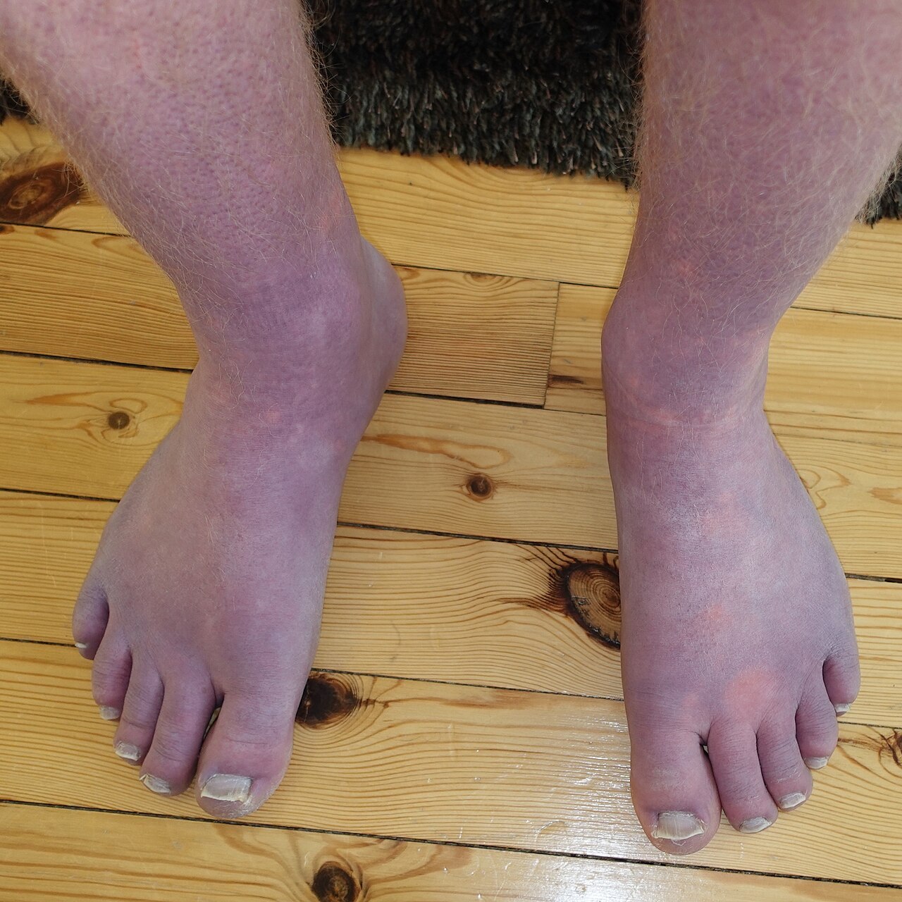
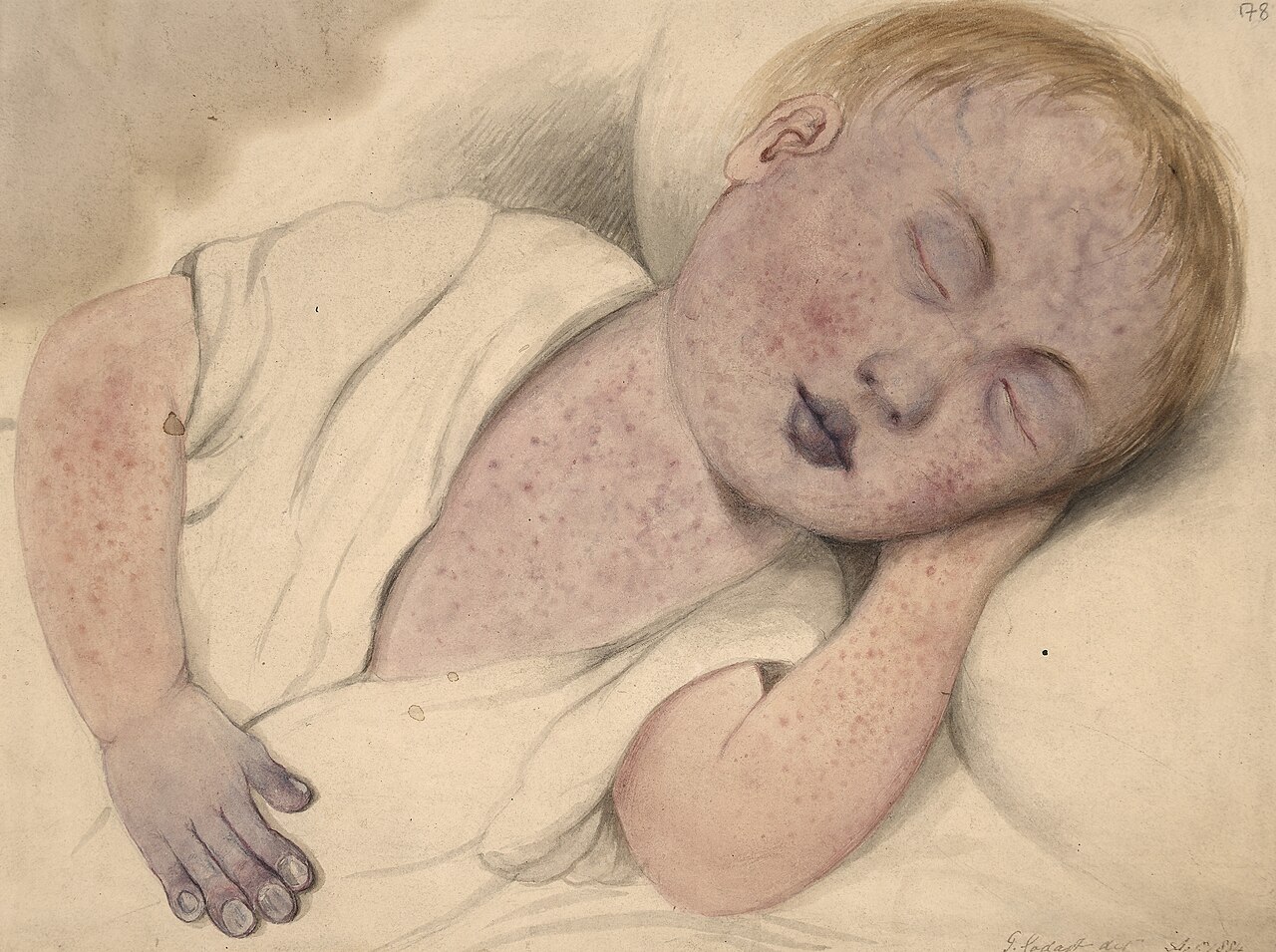
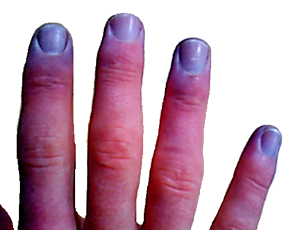
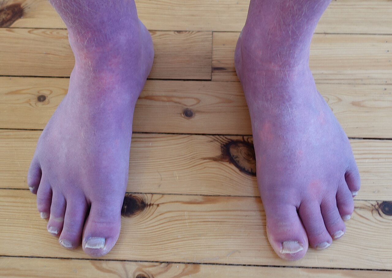
Cyanosis describes a bluish-purple discoloration of the skin and tissues, caused by a drop in the amount of oxygen attached to hemoglobin within the capillary bed's red blood cells. This symptom is most noticeable in areas where the skin is thin, such as the mucous membranes, lips, nail beds, and earlobes. Certain medications, including those containing amiodarone or silver, can also produce a similar discoloration. Additionally, Mongolian spots, large birthmarks, or foods with blue or purple dyes may be mistaken for cyanosis. A careful physical exam and patient history are essential for an accurate diagnosis. Treatment targets the underlying cause, as cyanosis itself is not a disease but a sign of another condition. It is divided into two main types: central and peripheral.
**Pathophysiology**
The process behind cyanosis differs depending on whether it is central or peripheral.
**Central cyanosis** Central cyanosis results from low arterial oxygen saturation (SaO₂). It becomes visible when the concentration of deoxygenated hemoglobin in the blood reaches at least 5.0 g/dL (≥ 3.1 mmol/L), which corresponds to an oxygen saturation of about 85% or less. This usually points to a heart or lung problem.
**Peripheral cyanosis** Peripheral cyanosis occurs when deoxygenated hemoglobin builds up on the venous side of the peripheral circulation. The condition depends on the concentration of deoxygenated hemoglobin, not just the total amount. For instance, people with severe anemia may not appear cyanotic even if their deoxygenated hemoglobin levels are relatively high, while those with excess red blood cells (such as in polycythemia vera) can look cyanotic with lower levels.
**Causes**
**Central cyanosis** Central cyanosis is typically caused by a circulatory or breathing problem that prevents proper oxygenation of blood in the lungs. It appears when arterial oxygen saturation falls below 85% or 75%. Acute cyanosis can result from asphyxiation or choking and is a clear sign that the airway is blocked.
Possible causes include: - **Central nervous system issues** that impair normal breathing: intracranial hemorrhage, drug overdose (e.g., heroin), or generalized tonic-clonic seizure. - **Respiratory system problems**: pneumonia, bronchiolitis, bronchospasm (e.g., asthma), pulmonary hypertension, pulmonary embolism, hypoventilation, or COPD (emphysema).
Quick Facts
- Field
- Pulmonology, cardiology, emergency medicine
- Symptoms
- Hypothermia, numbness in the area where the cyanosis is, coughing, wheezing, difficulty breathing
- Types
- Circumoral, central, peripheral
- Causes
- Airway problems, lung problems, heart problems, exposure to extreme cold
- Differential
- Circumoral cyanosis, peripheral cyanosis, central cyanosis
- Prevention
- Avoid exposure to freezing cold temperatures, limit smoking or caffeine, avoid touching cyanide
Facts from the source article.
Lore & Background
Cyanosis is apparent usually in body tissues covered with thin skin, including mucous membranes, lips, nail beds, and ear lobes. Some medications, such as those containing amiodarone or silver, may cause discoloration. Mongolian spots, large birthmarks, and consumption of food products with blue or purple dyes can also result in bluish skin discoloration and may be mistaken for cyanosis. Appropriate physical examination and history taking is crucial to diagnose cyanosis.
Reader's Guide
Cyanosis is a symptom, not a disease, so management focuses on treating the underlying cause. In emergencies, management begins with securing the airway, breathing, and circulation. Supplemental oxygen should be given immediately in cases of significant respiratory distress. If methemoglobin levels are positive, first-line treatment is methylene blue. The name cyanosis literally means 'the blue disease' or 'the blue condition,' derived from the Greek word for blue. It was first described in 1749 by Jean-Baptiste de Sénac, who concluded from an autopsy that cyanosis was caused by a heart defect mixing arterial and venous blood. In 1919, Dr. Christen Lundsgaard derived the concentration of deoxyhemoglobin (8 volumes per cent) that could cause cyanosis.
Did You Know?
- Cyanosis begins to show once the concentration of deoxyhemoglobin in the blood reaches ≥ 5.0 g/dL (≥ 3.1 mmol/L) or oxygen saturation ≤ 85%.
- Patients with severe anemia may appear normal despite higher-than-normal concentrations of deoxyhemoglobin.
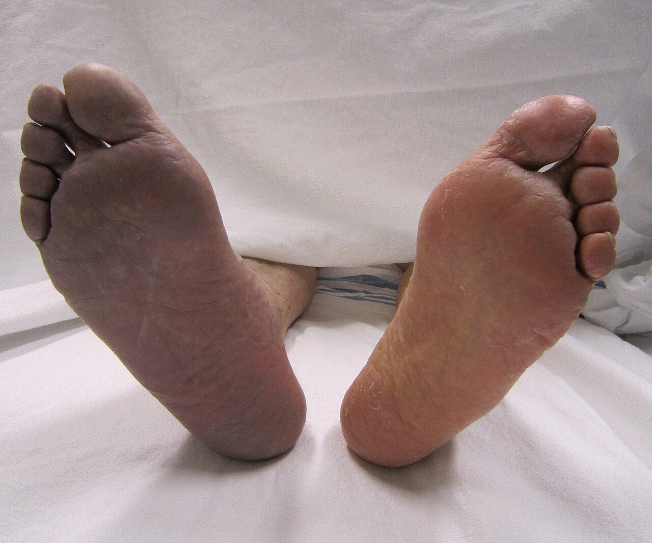
- Differential cyanosis, a bluish coloration of the lower but not the upper extremity and head, is seen in patients with a patent ductus arteriosus.
- Cyanosis may be more difficult to detect on people with darker skin pigmentation, but can still be diagnosed by examining nail beds, tongue, and mucous membranes.
The Mechanism Behind the Blue
Cyanosis manifests as a bluish-purple shift in tissue coloration, triggered when the oxygen bound to hemoglobin within the capillary bed falls below a critical threshold. This discoloration becomes visible primarily where skin is thin and highly vascularized—mucous membranes, the lips, nail beds, and earlobes. Clinicians divide the condition into two mechanistic categories. Central cyanosis arises when arterial oxygen saturation drops to 85 percent or lower, corresponding to a deoxyhemoglobin concentration of at least 5.0 grams per deciliter, and signals an underlying cardiopulmonary problem. Peripheral cyanosis, by contrast, reflects an elevated deoxyhemoglobin level on the venous side of the peripheral circulation, often without any heart or lung failure. Importantly, the visual appearance depends on total hemoglobin mass: a severely anemic patient may look entirely normal despite dangerously high deoxyhemoglobin levels, while someone with polycythemia vera can appear cyanotic at comparatively lower deoxyhemoglobin concentrations. A rare variant, differential cyanosis, produces a blue lower body with a pink upper body when a patent ductus arteriosus reverses its shunt direction.
The Many Roads to Cyanosis
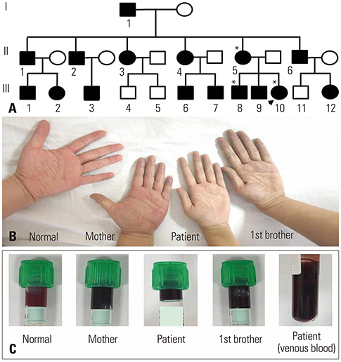
The triggers behind cyanosis span an extraordinary range of physiological disruptions. Central forms typically stem from circulatory or ventilatory failures that impair blood oxygenation in the lungs. Acute episodes can result from asphyxiation or choking, serving as a definitive sign that ventilation is blocked. Slower-onset causes include central nervous system events such as intracranial hemorrhage, opioid overdose, and generalized tonic-clonic seizures; respiratory conditions like pneumonia, bronchiolitis, bronchospasm, pulmonary embolism, and chronic obstructive pulmonary disease; and cardiovascular problems including congenital heart defects such as Tetralogy of Fallot, heart failure, valvular disease, and myocardial infarction. Rare hemoglobinopathies—methemoglobinemia, sulfhemoglobinemia, and a congenital variant called HbM Boston caused by a single amino acid substitution—can also produce the blue hue. Peripheral cyanosis, meanwhile, may arise from cold exposure, reduced cardiac output, arterial obstructions like Raynaud phenomenon or peripheral vascular disease, and venous obstructions such as deep vein thrombosis. High-altitude exposure above 2,400 meters, hypothermia, frostbite, and obstructive sleep apnea round out the list. Notably, certain medications containing amiodarone or silver, large birthmarks known as mongolian spots, and even blue or purple food dyes can mimic the condition.
Reading the Signs: Evaluation and Diagnosis
Because cyanosis is a visible symptom rather than a standalone disease, accurate diagnosis hinges on a thorough history and focused physical examination, particularly of the cardiopulmonary system. In newborns, peripheral cyanosis typically appears in the distal extremities, around the mouth, and in the periorbital region, while the mucous membranes stay pink—a key distinction from central cyanosis, where mucous membranes themselves turn blue. Skin pigmentation complicates visual assessment; in individuals with darker skin, the discoloration is harder to spot, though careful inspection of the nail beds, tongue, and mucous membranes—areas with thinner, more vascular tissue—can still reveal it. A confounding factor is severe anemia, which can mask cyanosis entirely despite elevated deoxyhemoglobin; clinicians must then look for alternative signs such as pale lips, eyelids, and gums, persistent fatigue, lightheadedness, and irregular heartbeats. Once the clinical picture is established, a battery of investigations guides further workup: pulse oximetry, arterial blood gas analysis, complete blood count, methemoglobin level measurement, electrocardiogram, echocardiogram, chest X-ray, CT scan, cardiac catheterization, and hemoglobin electrophoresis.
Treating the Symptom, Not the Sign
Because cyanosis is fundamentally a sign of an underlying problem rather than a disease in its own right, all management strategies center on identifying and correcting the root cause. In any emergency setting, the immediate priority follows the standard sequence of securing the airway, restoring adequate breathing, and stabilizing circulation. For patients in significant respiratory distress, supplemental oxygen is administered without delay, delivered through a nasal cannula for milder cases or continuous positive airway pressure for more severe presentations. When the blue discoloration stems from restricted small blood vessels, the therapeutic goal shifts toward restoring normal oxygenation levels in the blood, which can reverse the peripheral tint. For hemoglobinopathies such as methemoglobinemia, targeted pharmacologic intervention addresses the abnormal hemoglobin directly. In cases of congenital cardiac shunts, surgical or interventional correction of the structural defect becomes the definitive treatment. The overarching principle remains consistent: the clinician's task is not to treat the color itself but to resolve the cardiopulmonary, vascular, or metabolic disturbance that produced it, whether that disturbance is a pulmonary embolism, a drug-induced methemoglobinemia, a congenital heart defect, or a simple cold-induced vascular spasm.
Gallery
Frequently Asked Questions
Who is Cyanosis?
Cyanosis is a clinical sign in which the skin and underlying tissues take on a blue-purple hue because the red blood cells in the capillaries are carrying less oxygen than normal. It is not a disease in itself but a visible warning that oxygen delivery to the tissues is compromised.
Where does Cyanosis show up most clearly?
The discoloration is easiest to spot in regions with very thin overlying skin, including the lips, the inner lining of the mouth, the beds beneath the fingernails, and the earlobes. These areas let the color of the blood vessels beneath show through more readily than thicker skin elsewhere on the body.
What is Cyanosis's origin story?
The sign was first formally described in 1749 by the French physician Jean-Baptiste de Sénac, who documented the bluish tint associated with inadequate oxygenation. Since then it has become one of the most recognizable bedside observations in medicine.
What are Cyanosis's two forms and how do they differ?
Central cyanosis arises when arterial blood itself is poorly oxygenated, while peripheral cyanosis results from sluggish venous flow that lets deoxyhemoglobin build up in the extremities. Clinicians typically confirm the sign when deoxyhemoglobin reaches at least 5 g/dL or when measured oxygen saturation falls to 85 percent or below.
Who are Cyanosis's common lookalikes?
Several unrelated findings can mimic the blue-purple tint, including large congenital birthmarks, foods heavily dyed blue or purple, and certain drugs such as amiodarone-containing products or silver preparations. A thorough history and focused physical exam are needed to separate true cyanosis from these imitators.
More in Symptoms and signs: Skin and subcutaneous tissue 1-23
Spotted an error? Know more?
Reader corrections go straight into our review queue. Suggest an edit · How this site is sourced