
Ascites
Abnormal fluid accumulation in the peritoneal cavity, often from cirrhosis.

Netha Hussain · CC BY-SA 3.0
Ascites is the abnormal accumulation of fluid in the peritoneal cavity, defined as more than 25 milliliters of fluid. It is a common complication of liver cirrhosis and other conditions, often leading to abdominal distension, discomfort, and shortness of breath. Its management involves addressing the underlying cause and relieving symptoms through diet, medication, or fluid drainage.
Quick Facts
- Pronounce
- @ · ' · s · ai · t · i: · z, ə · SY · teez
- Field
- Gastroenterology, general surgery
- Symptoms
- Increased abdominal size, increased weight, abdominal discomfort, shortness of breath
- Complications
- Spontaneous bacterial peritonitis, hepatorenal syndrome, low blood sodium
- Causes
- Liver cirrhosis, cancer, heart failure, tuberculosis, pancreatitis, blockage of the hepatic vein
- Diagnosis
- Physical exam, ultrasound, CT scan
- Treatment
- Low-salt diet, medications, draining the fluid
- Medication
- Spironolactone, furosemide
- Frequency
- >50% of people with cirrhosis
Facts from the source article.
Lore & Background
Ascites derives from the Greek word 'askos,' meaning a leather bag or wineskin, reflecting the bag-like swelling of the abdomen. The condition is detected through physical signs such as flank bulging, shifting dullness, and fluid wave, and is graded from mild (visible only on imaging) to severe (directly visible). In the developed world, cirrhosis is the most common cause, with over half of cirrhosis patients developing ascites within ten years of diagnosis.
Reader's Guide
Ascites is a significant clinical condition because it indicates advanced liver disease or other serious underlying disorders. In cirrhosis, ascites arises from portal hypertension and vascular dysfunction, and its development carries a poor prognosis: half of those who develop ascites die within three years. Diagnosis relies on physical examination, imaging, and analysis of ascitic fluid, with the serum-ascites albumin gradient helping distinguish transudative from exudative causes. Treatment focuses on salt restriction, diuretics like spironolactone, and therapeutic paracentesis for severe cases. Refractory ascites, which does not respond to diuretics, may require a transjugular intrahepatic portosystemic shunt or liver transplant. Complications such as spontaneous bacterial peritonitis further increase morbidity. Understanding ascites is crucial for managing chronic liver disease and improving patient outcomes.
Did You Know?
- Ascites is defined as more than 25 milliliters of fluid in the peritoneal cavity.
- In the developed world, the most common cause of ascites is liver cirrhosis.
- Of those with cirrhosis who develop ascites, half will die within three years.
The Clinical Picture: From Subtle Swelling to Massive Distension
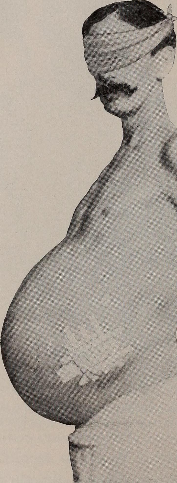
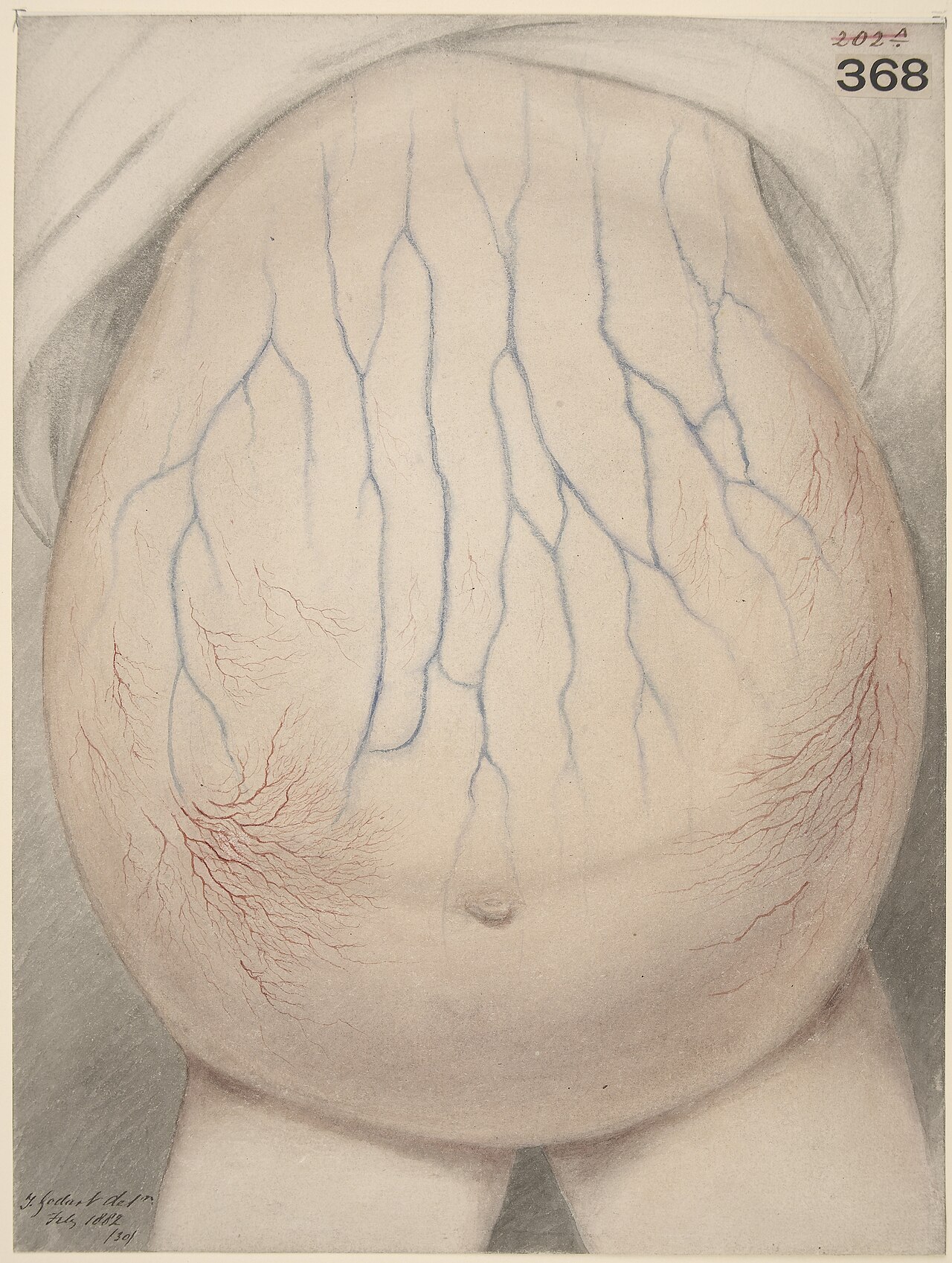
Ascites ranges from a barely perceptible accumulation of fluid to a dramatic distension of the abdomen capable of holding as much as thirty-five litres. In its mildest form, the condition is nearly invisible to the naked eye and only revealed by imaging. As volume grows, patients typically report a progressive sense of heaviness and pressure in the belly, along with breathlessness caused by the fluid pressing upward against the diaphragm.
At the bedside, clinicians rely on a handful of classic physical manoeuvres. In a reclining patient, fluid collects along the flanks, producing a visible bulge. Percussion notes shift when the person is rolled onto their side, a finding known as shifting dullness. In massive cases, tapping one side of the abdomen generates a palpable wave that travels across to the opposite flank.
Beyond the abdomen, the underlying aetiology stamps its own signature on the patient. Portal hypertension may bring leg oedema, easy bruising, gynecomastia, vomiting of blood, or the confusion of hepatic encephalopathy. Malignant ascites often accompanies chronic fatigue and weight loss, while cardiac ascites adds wheezing and exercise intolerance. A feared complication, spontaneous bacterial peritonitis, can strike at any stage.
Etiology and the SAAG Framework
Understanding why fluid accumulates in the peritoneal cavity hinges on a single laboratory measurement: the serum-ascites albumin gradient, or SAAG. A gradient above 1.1 g/dL points to portal hypertension as the driving force, while a value below that threshold signals a non-portal mechanism such as inflammation or malignancy.
Among portal-hypertensive causes, liver cirrhosis dominates, accounting for roughly 81 percent of cases. Within that group, alcoholic aetiology represents about 65 percent, viral hepatitis around 10 percent, and a cryptogenic or unexplained cause makes up the remaining 6 percent. Heart failure contributes approximately 3 percent. Rarer portal-hypertensive culprits include Budd-Chiari syndrome, veno-occlusive disease, constrictive pericarditis, and the childhood protein-energy malnutrition known as kwashiorkor.
On the low-gradient side, metastatic or primary peritoneal cancer accounts for about 10 percent of ascites. Tuberculosis and spontaneous bacterial peritonitis each represent roughly 2 percent, pancreatitis about 1 percent, and the remainder is spread across serositis, nephrotic syndrome, and hereditary angioedema. Mechanistically, transudates arise from elevated portal pressures—often around 20 mmHg—whereas exudates are actively secreted by inflamed or malignant tissue, carrying higher protein, lower pH, and more white cells.
Diagnostic Workup and Imaging
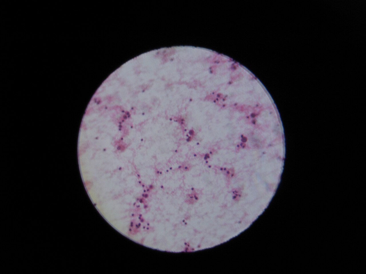
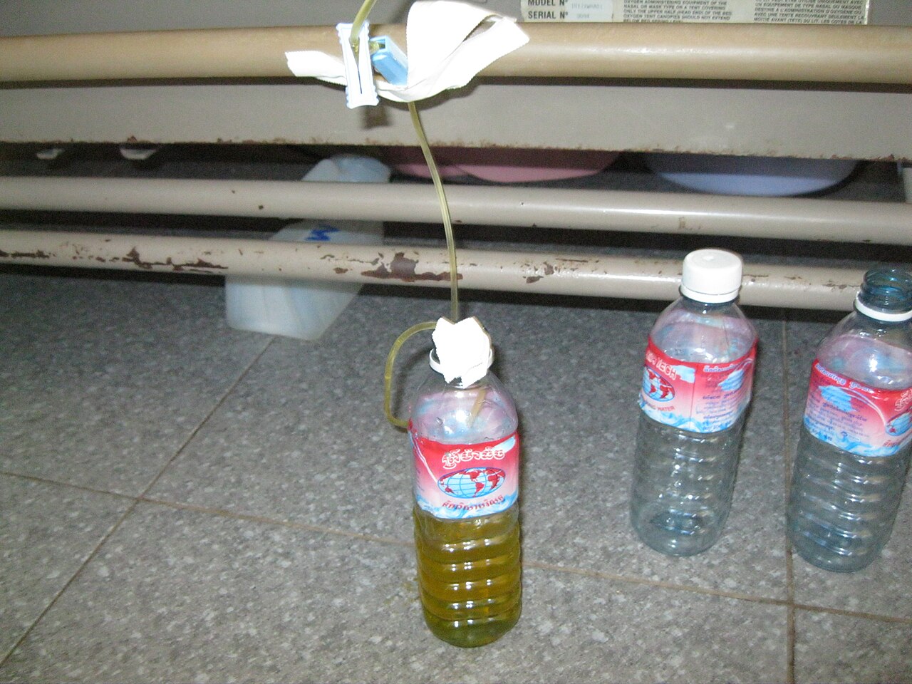
Once ascites is suspected, the diagnostic pathway begins with routine blood work—complete blood count, basic metabolic profile, liver enzymes, and coagulation studies. Most specialists advocate a diagnostic paracentesis whenever the fluid is newly detected or the patient is being admitted to hospital. The aspirated sample is examined for gross appearance, protein, albumin, and cell counts. Depending on context, microbiological culture, Gram stain, or cytopathology may follow.
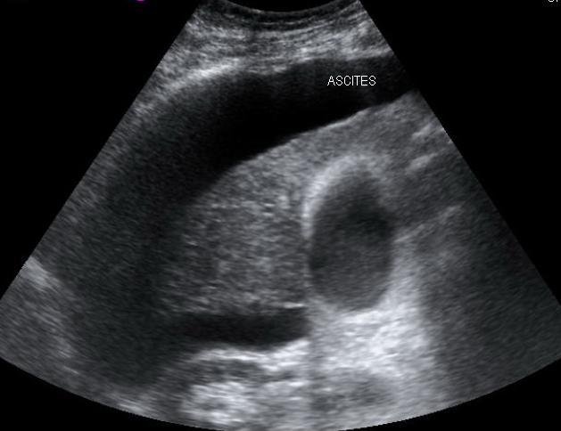
Imaging plays a complementary role. An ultrasound performed before any drainage attempt can reveal organ size and shape, estimate free-fluid volume, and, with Doppler studies, trace flow within the portal vein. It can also unmask Budd-Chiari syndrome or portal vein thrombosis. For difficult-to-drain collections, the sonographer may guide the needle in real time. When finer anatomical detail is needed, an abdominal CT scan offers superior resolution of organ structure compared with a sonogram.
Clinicians further stratify ascites into uncomplicated and refractory categories. Uncomplicated ascites—graded from ultrasound-only detection to a visible fluid wave—responds to salt restriction and diuretics. Refractory ascites, which recurs despite those measures, splits into diuretic-intractable and diuretic-resistant subtypes.
Treatment Pathways and Prognosis
Managing ascites is a layered effort that typically starts with dietary salt restriction and pharmacological diuresis, supplemented by periodic drainage of the accumulated fluid. For patients whose condition does not yield to these measures, a transjugular intrahepatic portosystemic shunt (TIPS) may be placed to relieve portal pressures, though the procedure carries its own complications. In the most severe cases, treating the root cause through a liver transplant becomes the definitive option.
The prognosis, however, remains sobering. Among patients diagnosed with cirrhosis, more than half will develop ascites within ten years. Of that subset, roughly half will die within three years of the fluid first appearing, underscoring how ascites marks a critical inflection point in the natural history of liver disease.
Refractory ascites, the form that persists or returns despite adequate diuretic therapy and salt restriction, is the most challenging variant. It divides into two subtypes: diuretic-intractable ascites, where the drugs cannot be escalated because of side effects such as rising creatinine or potassium depletion, and diuretic-resistant ascites, where the fluid simply does not respond to the medication. This distinction shapes the urgency with which clinicians consider shunt placement or transplantation.
Gallery
Frequently Asked Questions
What is Ascites in the context of digestive system signs?
Ascites refers to an abnormal buildup of fluid within the peritoneal cavity, specifically when the volume exceeds 25 milliliters. It is most frequently linked to liver cirrhosis, though other conditions can also trigger it.
What are the main symptoms and signs of Ascites?
Patients typically present with noticeable abdominal distension, a sense of discomfort, and shortness of breath. These signs stem from the pressure the excess fluid exerts on surrounding organs.
How is Ascites diagnosed?
Clinicians rely on a combination of physical examination, ultrasound, and CT imaging to confirm fluid presence. The serum-ascites albumin gradient (SAAG) helps determine whether the underlying driver is a high-gradient cause like cirrhosis.
What is the most common underlying cause of Ascites?
Liver cirrhosis accounts for roughly 81% of high-gradient ascites cases, making it by far the leading association. Other etiologies exist, but cirrhosis dominates the clinical picture.
How is Ascites managed and treated?
Treatment centers on addressing the root cause while alleviating symptoms through a low-salt diet, diuretic medications, and therapeutic fluid drainage (paracentesis). In more severe or refractory cases, options include TIPS placement or liver transplantation.
More in Symptoms and signs: Digestive system and abdomen 1-24
Spotted an error? Know more?
Reader corrections go straight into our review queue. Suggest an edit · How this site is sourced