Decubitus ulcer
Localized skin damage from prolonged pressure, often over bony prominences.
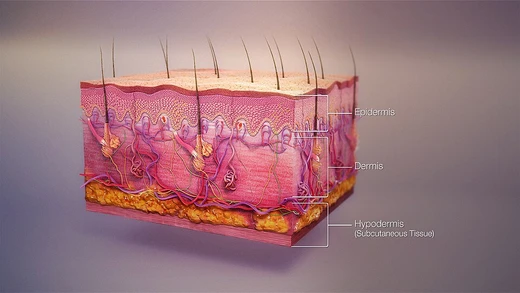
Decubitus ulcers, also called pressure ulcers or bed sores, are localized injuries to the skin and underlying tissue, typically over bony prominences, caused by prolonged pressure, shear, or friction. They are a significant concern in healthcare settings due to their high prevalence and potential for serious complications, including death.
Quick Facts
- Prevalence in european hospitals
- 8.3% to 23%
Facts from the source article.
Did You Know?
- Paralyzed individuals are the most likely to have pressure sores recur.
- The most common causes of fatality from pressure ulcers are kidney failure and amyloidosis.
Pathophysiology
Pressure ulcers arise from inadequate blood supply to tissues, leading to ischemia and reperfusion injury when blood flow returns. A mild example occurs in healthy individuals sitting still for long periods, where a dull ache signals impeded blood flow; within two hours, ischemia can cause tissue damage and cell death, initially presenting as a red, painful area. Alternatively, high pressure can directly damage muscle cell membranes, causing muscle death and subsequent skin death through compromised blood vessels, a deep tissue injury form that begins as purple intact skin. Biofilm, which forms rapidly in wounds, is a common cause of delayed healing by maintaining inflammation; it requires frequent debridement and antimicrobial dressings. Infection also prevents healing, indicated by slow healing, pale granulation tissue, and systemic signs such as fever, pain, redness, swelling, warmth, and purulent discharge; infected wounds may have a gangrenous smell, discoloration, and increased pus.
Diagnosis
Pressure ulcer stages are defined by the National Pressure Injury Advisory Panel in the United States and the European Pressure Ulcer Advisory Panel in Europe, with revisions made periodically. Stage 1 involves intact skin with non-blanchable redness over a bony prominence; in darkly pigmented skin, blanching may not be visible, and the area may differ in color, thickness, or temperature. Stage 2 is partial-thickness dermis loss, appearing as a shallow open ulcer with a red-pink bed without slough, or as an intact or ruptured serum-filled blister; this stage excludes skin tears, tape burns, perineal dermatitis, maceration, or excoriation. Stage 3 involves full-thickness tissue loss with visible subcutaneous fat but no exposed bone, tendon, or muscle; slough may be present, and depth varies by location—shallow on areas like the nose or ear, deep where adiposity is significant. Stage 4 is full-thickness loss with exposed bone, tendon, or muscle; slough or eschar may be present, and undermining or tunneling is common; osteomyelitis is likely, and exposed cartilage is also classified as stage 4.
Prevention
Prevention strategies include modifications to bedding and mattresses, support systems to relieve pressure, skin care, nutrition, and organizational changes in care routines. The Standardized Pressure Injury Prevention Protocol (SPIPP) Checklist, derived from the International Guideline, was established as law for United States Department of Veterans Affairs facilities in 2022 to ensure patient safety. Before repositioning, a risk assessment tool such as the Braden Scale, Norton, or Waterlow is recommended to determine the best approach; these tools score factors like moisture, activity, and mobility to create an individualized plan for turning and repositioning. However, unbiased clinical studies are needed to determine the most effective interventions, and consistent implementation in hospitals is not always achieved.
Treatment
Treatment recommendations include bed rest, pressure-redistributing support surfaces, nutritional support, repositioning, wound care such as debridement and dressings, and biophysical agents like electrical stimulation, though reliable scientific evidence for many interventions is lacking. Necrotic tissue should be removed in most cases, except on the heel when blood supply is inadequate, using five methods: autolytic debridement with moist dressings, biological debridement with medical maggots (approved by the FDA in 2004), chemical debridement with prescribed enzymes, mechanical debridement with dressings or ultrasound, and surgical debridement for fastest removal. It is unclear if one topical agent or dressing is superior; some evidence suggests protease-modulating dressings, foam dressings, or collagenase ointment may be better than gauze, and antimicrobial products may improve healing, but no international consensus exists on dressing selection.
More in Skin Conditions Resulting from Physical Factors
Spotted an error? Know more?
Reader corrections go straight into our review queue. Suggest an edit · How this site is sourced