Coma blister
Pressure-induced blisters in comatose individuals, often from drug overdose.
A coma blister, or coma bullae, is a skin lesion that arises from pressure in individuals with impaired consciousness. These blisters, which can be hemorrhagic and range from 4 to 5 centimeters in diameter, are most often found on the extremities and trunk. They are notable for their association with drug overdose, particularly of central nervous system depressants, though they also occur in various metabolic and neurologic conditions.
Quick Facts
- Size range
- 4 to 5 centimeters in diameter
- Common locations
- Extremities and trunk
- Associated drug classes
- Barbiturates
- tricyclic antidepressants
- hypnotics
- benzodiazepines
- opiates
- antipsychotics
- alcohol
- Frequency in barbiturate overdose
- 4%
- Typical onset after unconsciousness
- 48 to 72 hours
- Typical healing time
- One to two weeks
- More common in
- Adults
Facts from the source article.
Etiology
Coma blisters develop from inadequate oxygen at the tissue level combined with external pressure, often in the setting of neurologic disease or metabolic disorders. Neurologic conditions such as cranioencephalic trauma, cerebrovascular disease, and meningoencephalitis are associated with their formation, as are metabolic disturbances like diabetic ketoacidosis, hyperkalemia, and hypoglycemia. Drug overdose-induced comas are a frequent predisposing factor, with studies reporting blisters in 4% of barbiturate overdoses. Recent research suggests immune mechanisms may play a role, particularly in drug-induced comas, where direct immunofluorescence staining reveals IgG, IgA, and C3 with fibrin. A common feature across causes is necrosis of the eccrine sweat glands. Constant pressure from immobilization injures vessel walls, interrupting blood and oxygen flow to tissues, leading to necrotic bullae and deterioration of sweat glands. This pressure is often exacerbated by vasoactive drugs or shock, which lower arterial blood pressure and trigger a reflex that increases pressure on arterial walls. Blisters typically form on pressure points such as the ankles, heels, knees, elbows, fingers, and toes. Administration of antidepressants, antipsychotics, barbiturates, benzodiazepines, ethanol, or opioids is linked to overdose-induced comas due to their vasoactive properties and potential toxicity to eccrine sweat glands. Blisters appear at vasoactive pressure sites 48 to 72 hours after the onset of unconsciousness.
Diagnosis
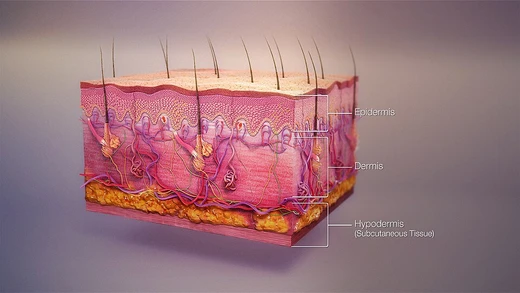
Coma blisters usually develop on pressure point sites within a few days in individuals immobilized due to external events, with barbiturate overdose being the most frequent predisposing event. Any condition that renders a person unconscious can lead to their formation. The blisters typically resolve on their own within one to two weeks. Histological evidence aids diagnosis and includes subepidermal bullae, focal necrosis of the epidermis, dermis, subcutaneous tissue, and all epidermal appendages. In non-drug-induced coma blisters, the absence of inflammation and the presence of thrombosis in dermal wall vessels are the two most significant differences from drug overdose-induced cases. Necrosis of sweat glands and ducts is common but not required for diagnosis. In children, careful clinicopathological correlation is needed to exclude other blistering diseases. Friction blisters differ as they occur only in areas of repetitive friction, caused by mechanical separation of epidermal cells at the stratum spinosum, with fluid filling the separation due to hydrostatic pressure. Edema blisters form at sites of swelling from acute volume overload, such as from renal or heart failure, and histopathology may show subepidermal edema with negative immunofluorescence staining. Bullous diabeticorum occurs in individuals with diabetes mellitus, often with nephropathy and neuropathy, and may result from premature aging of the local subbasement membrane zone connective tissue. Epidermolysis bullosa is a genetic disease causing extreme skin fragility and blistering from minimal friction or trauma, with no cure and treatment focused on wound management.
Treatment and management
Coma blisters typically do not require treatment and heal on their own within one to two weeks. Early identification and management involve treating any underlying conditions that led to blister formation and reducing the risk of subsequent infections. Before starting treatment, other potential bullous diseases such as epidermolysis bullosa, bullous pemphigoid, bullous amyloidosis, and epidermolysis bullosa acquisita should be ruled out, as should friction blisters and edema blisters. If treatment is needed, topical antibiotics can prevent infection. Management and wound care may include sterile drainage of the blister, positioning the individual away from the wound to relieve pressure, and preventing pressure ulcerations. When draining, leaving the blister roof intact allows it to act as an additional dressing layer, and hydrocolloid dressings can maintain moisture and promote healing. Some individuals may experience visible scarring after healing, but this is non-concerning. Coma blisters are not contraindications for other medications or therapies, so medications for co-morbidities should be continued as needed.
More in Skin Conditions Resulting from Physical Factors
Spotted an error? Know more?
Reader corrections go straight into our review queue. Suggest an edit · How this site is sourced