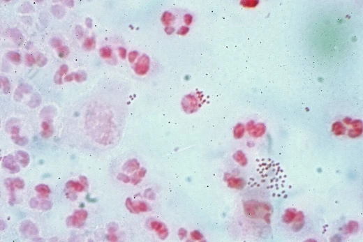
Mycoplasma pneumoniae
Smallest self-replicating bacterium causing atypical pneumonia in humans.

Mycoplasma pneumoniae is a bacterium, but an unusual one. It belongs to a class called Mollicutes, and its cells are extremely small and completely lack a cell wall. In humans, it is the cause of Mycoplasma pneumonia, a type of atypical bacterial pneumonia that is also linked to cold agglutinin disease. It ranks among the tiniest organisms capable of replicating on their own.
The story of its discovery began in 1898, when researchers Nocard and Roux isolated a microbe from cattle with pneumonia. This microbe resembled other organisms soon found in various animals, known as pleuropneumonia-like organisms (PPLOs), which were later tied to pneumonias and arthritis in those species. A major step forward came in 1944, when Monroe Eaton grew an agent from human pneumonia cases inside embryonated chicken eggs; this became known as the "Eaton agent." Because it could be cultivated in eggs (a method then used for viruses) and because antibiotics worked against the infection, its true nature was unclear. In 1961, NIH researcher Robert Chanock, who was studying the Eaton agent, visited Leonard Hayflick at the Wistar Institute. Chanock suspected the agent might not be a virus. Hayflick, whose doctoral work had involved PPLOs in animals, suggested it could be a mycoplasma. Chanock sent him egg yolk containing the agent, and Hayflick, using a new culture medium he had developed, isolated a unique mycoplasma. Chanock and Hayflick then proved this organism caused human pneumonia. The leading expert on these organisms, Emmy Klieneberger-Nobel, proposed naming it *Mycoplasma hayflickiae*, but Hayflick declined, and it was instead named *Mycoplasma pneumoniae*.
Taxonomically, *M. pneumoniae* sits within the Mollicutes, a group defined by the absence of a peptidoglycan cell wall. This lack makes them naturally resistant to antibiotics like beta-lactams that target cell wall construction. Mycoplasmas have a stripped-down genome and simple metabolism, making them obligate parasites that depend on their host for many resources. *M. pneumoniae* uses a specialized attachment organelle to stick to cells in the respiratory tract, which also helps it move and invade. Its ability to persist after treatment is partly due to its mimicking of host cell surface molecules.
The bacterium causes disease by adhering to host cells and releasing cytotoxic agents. This leads to loss of cilia and the release of hydrogen peroxide, driving respiratory symptoms and complications like bronchial asthma and chronic obstructive pulmonary disease. It also produces a unique toxin called CARDS, which fuels inflammation and breathing problems. Treatment usually involves macrolides or tetracyclines, which stop protein synthesis. However, resistance—especially in Asia—is on the rise, mostly from mutations in the 23S rRNA gene that block macrolide binding, making management harder and requiring alternative therapies.
- field
- Microbiology
- known_for
- Causing atypical pneumonia (walking pneumonia); first mycoplasma proven to cause human disease
Quick Facts
- Genus
- Mycoplasmoides
- Species
- pneumoniae
Facts from the source article.
Lore & Background
These PPLOs were soon linked to pneumonias and arthritis in several animals. Hayflick, whose doctoral dissertation had been on animal diseases caused by PPLOs, reasoned the Eaton agent might be a mycoplasma. Using a novel agar and fluid medium he devised, Hayflick isolated a unique mycoplasma from egg yolk containing the Eaton agent. This was soon proven by Chanock and Hayflick to be the causative agent of primary atypical pneumonia. Emmy Klieneberger-Nobel suggested naming it Mycoplasma hayflickiae, but Hayflick demurred in favor of Mycoplasma pneumoniae. Taxonomically, M. pneumoniae is part of the Mollicutes class, characterized by lack of a peptidoglycan cell wall, making it inherently resistant to beta-lactam antibiotics. It uses a specialized attachment organelle to adhere to respiratory tract cells, and its persistence is associated with its ability to mimic host cell surface composition.
Reader's Guide
Mycoplasma pneumoniae holds significant importance in medical microbiology as the first mycoplasma proven to cause a human disease. The organism's lack of a cell wall makes it inherently resistant to antibiotics targeting cell wall synthesis, such as beta-lactams, and treatment typically involves macrolides or tetracyclines that inhibit protein synthesis. However, resistance has been increasing, particularly in Asia, due to mutations in the 23S rRNA gene. Pathogenic mechanisms include host cell adhesion, cilia loss, hydrogen peroxide release, and production of a unique CARDS toxin, contributing to respiratory symptoms and complications such as bronchial asthma and chronic obstructive pulmonary disease. The organism's reduced genome and dependence on host metabolism highlight its parasitic lifestyle. Its discovery also advanced understanding of mycoplasmas as a class, which are among the smallest self-replicating organisms and are thought to have evolved by degenerative evolution from gram-positive eubacteria. The inverted microscope under which Hayflick discovered M. pneumoniae is kept by the Smithsonian Institution.
Did You Know?
- Mycoplasma pneumoniae is one of the smallest self-replicating organisms, with cells approximately 0.1–0.2 μm wide and 1–2 μm long.
- The organism lacks a cell wall, making it inherently resistant to beta-lactam antibiotics like penicillin.
Frequently Asked Questions
Who is Mycoplasma pneumoniae?
Mycoplasma pneumoniae is a tiny, wall-less bacterium in the class Mollicutes and the first member of the mycoplasma group ever confirmed as a human pathogen. It ranks among the smallest organisms on Earth that can still replicate on its own.
What are Mycoplasma pneumoniae's powers or role?
Its signature move is triggering atypical pneumonia—widely nicknamed 'walking pneumonia'—and it can also be tied to cold agglutinin disease. Because it lacks a cell wall entirely, it dodges the whole class of antibiotics that work by disrupting wall synthesis.
How does Mycoplasma pneumoniae's story end?
In a typical infection the host's immune system eventually clears the organism, although the lingering cough and respiratory symptoms can stretch on for several weeks. There is no chronic 'villain arc'; once eliminated, it does not establish a lifelong residency in the body.
More in Pathogenic Bacteria 1-20
Spotted an error? Know more?
This is a living reference — every entry is fact-audited, and reader corrections feed straight into our audit queue. Suggest an edit · See this site's audit record