Mycobacterium tuberculosis

Mycobacterium tuberculosis, often called Koch’s bacillus, is the bacterium that causes tuberculosis. It belongs to the Mycobacteriaceae family and primarily targets the lungs of mammals, needing a lot of oxygen to thrive. Its cell surface has an unusual waxy coating, largely made of mycolic acid, which prevents it from taking up Gram stain—so under that method it looks only weakly Gram-positive. Instead, scientists use acid-fast stains like Ziehl–Neelsen or fluorescent stains like auramine to spot it under a microscope. Robert Koch first identified this pathogen in 1882. Common diagnostic tools include the tuberculin skin test, acid-fast staining, culture, and polymerase chain reaction. The bacterium’s genome was fully sequenced in 1998.
Microbiologically, M. tuberculosis is nonmotile and strictly aerobic. It divides very slowly, once every 18 to 24 hours—contrast that with Escherichia coli, which can split in about 20 minutes. The cells are small, curved rods that can survive weak disinfectants and remain viable for weeks in dry conditions. Their lipid-rich cell wall, containing mycolic acid and cord factor glycolipid, helps them resist drying out and is a major factor in their ability to cause disease.
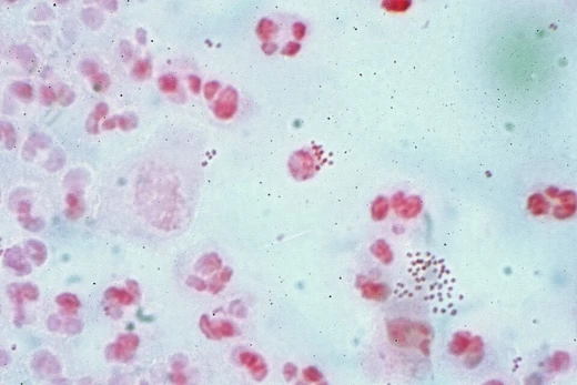
Under the microscope, M. tuberculosis cells don’t absorb Gram stain because of the mycolic acid. Instead, they’re visualized with acid-fast or fluorescent stains. The bacteria are curved rods that often clump together, thanks to fatty acids in their walls, forming rope-like strands called cording. In infected tissue, they produce caseating granulomas that contain Langhans giant cells, which have a characteristic horseshoe-shaped arrangement of nuclei.
Culturing M. tuberculosis in the lab is slow. It doubles about once a day. Common growth media include liquid types like Middlebrook 7H9 or 7H12, egg-based solids like Lowenstein-Jensen, and agar-based solids like Middlebrook 7H11 or 7H10. Visible colonies take several weeks to appear on agar plates. Special tubes containing a fluorescent gel can signal mycobacterial growth. To distinguish it from other mycobacteria, scientists test for catalase and niacin production, and confirm identity with gene probes or MALDI-TOF.
Scanning electron microscopy reveals that M. tuberculosis cells average 2.71 micrometers in length and 0.345 micrometers in diameter. The outer membrane surface area is about 3.04 square micrometers, and the plasma membrane surface area about 2.67 square micrometers. The cell’s total volume is roughly 0.293 femtoliters, broken down into outer membrane (0.006 fl), periplasm (0.060 fl), plasma membrane (0.019 fl), and cytoplasm (0.210 fl). On average, each cell contains about 1,672 ribosomes, with a density of around 716.5 ribosomes per 0.1 femtoliter.
M. tuberculosis belongs to a genetically related group of at least nine species: M. tuberculosis sensu stricto, M. africanum, M. canettii, M. bovis, M. caprae, M. microti, M. pinnipedii, M. mungi, and M. orygis.
Humans are the only known natural hosts for M. tuberculosis. Contrary to common belief, it isn’t spread by handshakes, toilet seats, shared food or drink, or toothbrushes. Instead, transmission happens through airborne droplets released when an infected person coughs, sneezes, speaks, or sings. Once inhaled into the lungs, the bacteria are engulfed by alveolar macrophages, but the macrophages can’t kill them. The bacterial cell wall contains cord factor glycolipids that block the fusion of the phagosome with the lysosome—the compartment full of antibacterial compounds. Specifically, M. tuberculosis interferes with the bridging molecule EEA1, though it still allows nutrient-filled vesicles to fuse. It also produces a diterpene called isotuberculosinol that stops phagosome maturation, and it neutralizes reactive nitrogen intermediates to evade macrophage killing. More recently, researchers found that the bacterium secretes and coats itself in 1-tuberculosinyladenosine (1-TbAd), a special nucleoside that acts like an antacid, neutralizing pH and causing lysosomes to swell.
In infections, levels of the protein PPM1A increase. This protein is involved in both intrinsic and extrinsic apoptotic pathways, and its elevated expression inhibits those pathways, preventing macrophages from undergoing normal apoptosis. Through kinome analysis, the JNK/AP-1 signaling pathway was identified as a downstream target of PPM1A’s effect on macrophage apoptosis. By suppressing apoptosis, M. tuberculosis creates a safe place to replicate and can maintain a latent state for long periods. Granulomas—organized clusters of immune cells—are a hallmark of tuberculosis. They play a dual role: they help regulate the immune response and limit tissue damage, but they can also help the infection spread. The ability to create M. tuberculosis mutants and test them individually has advanced understanding of these processes.
- discoverer
- Robert Koch
- field
- Microbiology
- type
- Pathogenic bacterium
- known_for
- Causing tuberculosis
Lore & Background
Mycobacterium tuberculosis is a small, nonmotile bacillus that divides extremely slowly, every 18–24 hours, compared to bacteria like Escherichia coli which divide roughly every 20 minutes. Its cell wall is rich in lipids such as mycolic acid and cord factor glycolipid, which contribute to its resistance to desiccation and weak disinfectants, and allow it to survive in a dry state for weeks. Under a microscope, cells are curved rod-shaped and often seen wrapped together in a pattern called cording, due to fatty acids in the cell wall. In tissue, it is characterized by caseating granulomas containing Langhans giant cells with a 'horseshoe' pattern of nuclei.
Reader's Guide
Mycobacterium tuberculosis is significant as the primary cause of tuberculosis, a major infectious disease that primarily affects the lungs. Its unusual cell wall, rich in mycolic acid, makes it resistant to Gram staining and requires specialized acid-fast stains like Ziehl–Neelsen or fluorescent stains like auramine for microscopic identification. The bacterium's slow growth rate—doubling roughly once per day—complicates laboratory culture, with visible colonies requiring several weeks on agar plates. Humans are the only known reservoirs, and transmission occurs through airborne droplets from coughing, sneezing, speaking, or singing. Inside the lungs, M. tuberculosis is phagocytosed by alveolar macrophages but evades killing by blocking phagosome-lysosome fusion, neutralizing reactive nitrogen intermediates, and secreting a nucleoside that neutralizes pH. The bacterium's ability to develop resistance to multiple drugs, due to gene mutations, poses ongoing challenges for treatment.
Did You Know?
- M. tuberculosis divides every 18–24 hours, extremely slow compared to Escherichia coli which divides roughly every 20 minutes.
- Its cell wall contains cord factor glycolipid, which inhibits fusion of the phagosome with the lysosome in macrophages.
- The bacterium can survive in a dry state for weeks and withstand weak disinfectants.
- Strain typing using variable numbers of tandem repeats (VNTR) helps investigate tuberculosis outbreaks.
Frequently Asked Questions
Who is Mycobacterium tuberculosis?
Mycobacterium tuberculosis, commonly nicknamed Koch's bacillus, is a pathogenic bacterium in the Mycobacteriaceae family and the sole organism responsible for causing tuberculosis. It is a strict respiratory pathogen that targets the lung tissue of mammals.
What are Mycobacterium tuberculosis's powers or special abilities?
Its signature trait is a thick, waxy outer layer packed with mycolic acid, which renders it nearly impossible to stain with a standard Gram procedure and gives it only a faint Gram-positive appearance. That same lipid-rich coat helps it shrug off many disinfectants and immune attacks, while its demand for high oxygen levels keeps it anchored in well-ventilated lung tissue.
How does Mycobacterium tuberculosis's story end?
In the canonical arc, a multi-drug antibiotic regimen—typically isoniazid, rifampin, ethambutol, and pyrazinamide—over six to nine months eradicates the organism from the patient. Without treatment, however, the bacterium can enter a long dormant phase and reactivate years later, making its 'ending' one of the most protracted storylines in all of microbiology.
More in Pathogenic Bacteria 1-20
Spotted an error? Know more?
This is a living reference — every entry is fact-audited, and reader corrections feed straight into our audit queue. Suggest an edit · See this site's audit record