
Degloving
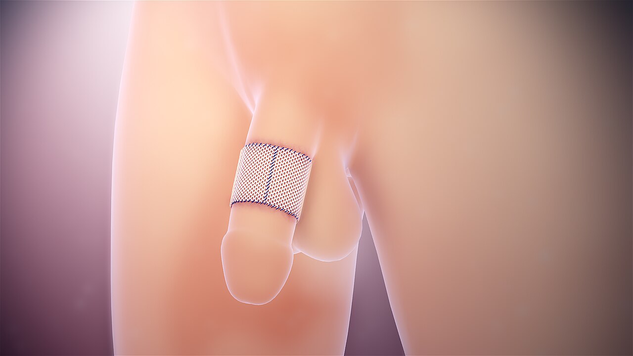
Skin and fat torn from underlying structures by shearing forces.

www.scientificanimations.com · CC BY-SA 4.0
Degloving is a type of avulsion injury in which the skin and underlying subcutaneous tissue are torn away from the deep fascia and muscle. It can occur anywhere on the body, commonly affecting the face, scalp, trunk, limbs, and genitalia, and is caused by shearing forces. Degloving injuries were first reported in the twentieth century from machinery such as clothes wringers, and later from automobile accidents.
- Type
- injury
- First reported
- twentieth century
- Common causes
- machinery, automobiles
- Affected areas
- face, scalp, trunk, limbs, genitalia
- Categories
- open, closed
Lore & Background
Degloving injuries are categorized as open or closed. In open injuries, the skin is torn back, exposing underlying structures, resembling the removal of a glove. Closed injuries, such as the Morel-Lavallée lesion, are internal separations between soft tissue layers that create a pocket that can fill with blood and lymphatic fluid. The Morel-Lavallée lesion is named after a French physician who first described it.
Reader's Guide
Degloving injuries represent a significant class of soft-tissue trauma, with treatment requiring careful assessment of tissue viability and blood supply. Dead tissue must be removed; healthy torn tissue with blood supply may be reattached via replantation or revascularization. When reattachment is not possible, skin flaps or grafting are used. The Morel-Lavallée lesion, a closed internal degloving injury, can be challenging to diagnose because external appearance may be normal despite underlying damage. It can lead to complications such as hypovolemic shock, skin necrosis, or infection. Diagnosis often relies on imaging, with MRI providing detailed views and the Mellado-Bencardino classification describing six types based on MRI features. Treatment aims to close the space between fascia layers, with conservative therapy for small fluid collections without capsule formation.
Did You Know?
- A closed internal degloving injury is called a Morel-Lavallée lesion, named after a French physician.
- Morel-Lavallée lesions can occur after liposuction or abdominoplasty.
- The Mellado-Bencardino classification categorizes Morel-Lavallée lesions into six types based on MRI features.
Anatomy and the Shearing Mechanism
Degloving is a form of avulsion in which the skin and the subcutaneous fat layer beneath it are forcibly stripped away from the deeper anatomical structures to which they are normally anchored. Under healthy conditions, the subcutaneous tissue rests against the deep fascia, a fibrous membrane that sheathes the muscles below. When a powerful shearing force is applied, the soft-tissue layers are pulled apart along this interface, separating the skin and fat from the muscle and fascia. This mechanism can produce injury at virtually any site on the body, though the face, scalp, trunk, limbs, and genitalia are frequently affected. The severity of the tear depends on the magnitude and direction of the force, and the resulting wound may range from a partial detachment to a complete separation of the soft-tissue envelope. Because the injury disrupts the normal attachment between the superficial and deep layers, the blood and lymphatic vessels running through the subcutaneous plane are often damaged, setting the stage for fluid accumulation and further tissue compromise.
Historical Context and Modern Causes
The first documented reports of degloving injuries emerged in the twentieth century, linked to industrial machinery such as clothes wringers that could catch and tear skin from the body. The subsequent proliferation of automobiles introduced an entirely new class of high-energy trauma, and motor vehicle collisions remain a leading cause of these wounds today. Beyond crashes, degloving can result from crush injuries, blunt-force impacts at lower velocities like falls, bicycle accidents, and sports-related trauma. Even in the operating room, procedures such as liposuction and abdominoplasty have been associated with post-operative degloving. The common thread across all these scenarios is a shearing force sufficient to overcome the natural attachment between the subcutaneous tissue and the deep fascia. The injury is not confined to a single region; it can manifest on the face, scalp, trunk, limbs, or genitalia, making it a concern for trauma surgeons, plastic surgeons, and emergency physicians alike.
Classification and Clinical Presentation
Degloving injuries fall into two broad categories based on whether the wound is open to the outside. In an open degloving, the skin is peeled back far enough that the deeper structures become visible, an appearance that literally evokes the act of pulling a glove off a hand. A closed degloving, by contrast, leaves the skin intact on the surface while the internal layers have separated, creating a sealed pocket between the superficial and deep fascia. The eponymous Morel-Lavallée lesion, named for a French physician who first described it, is the classic example of this closed variant. Clinically, the affected area may display bruising, swelling, cracking, drying, or abrasions, and the skin can feel abnormally mobile because it is no longer anchored to the muscle beneath. Fluid from damaged blood and lymphatic vessels collects in the new space, and in severe cases this can lead to hypovolemic shock, skin necrosis from pressure, or infection. In chronic presentations, a fibrous capsule forms inside the pocket, trapping fluid and preventing natural resorption. Adults most commonly develop these lesions at the hip and thigh, while children tend to present below the knee.
Diagnosis and Surgical Management
Recognizing a degloving injury can be straightforward or deceptively difficult. In polytrauma patients, life-threatening wounds demand immediate attention and may mask the subtler signs of a closed degloving, delaying diagnosis. When the external appearance is unremarkable, medical imaging becomes essential. Computed tomography is frequently the first modality employed because it is often obtained as part of the initial trauma workup, while magnetic resonance imaging provides the most detailed delineation of the fluid-filled space and surrounding soft tissue. Ultrasound also plays a role in the diagnostic toolkit. Treatment begins with a careful assessment of the soft-tissue damage and the status of associated blood vessels. Any tissue that has lost its viability must be excised. If the avulsed tissue remains healthy and retains an adequate blood supply, surgeons can perform replantation and revascularization to reattach it with restored circulation. When reattachment is not feasible, reconstructive options such as skin flaps or skin grafting are employed to close the defect and restore coverage.
Gallery
Frequently Asked Questions
Who is Degloving?
Degloving is a traumatic avulsion wound in which the skin and the fat layer beneath it get ripped free from the deeper fascia and muscle. It is classified as both an open and a closed injury type, depending on how the tissue separates.
What are Degloving's powers/role?
The driving mechanism is a shearing force that peels the outer skin and subcutaneous tissue away from the structures underneath. It can strike virtually any body region, with the face, scalp, torso, limbs, and genital area being the most commonly hit.
Why is Degloving important?
Degloving is a major concern in trauma care because it can involve large surface areas across the face, trunk, or extremities in a single event. Its dual open-and-closed classification means it can look deceptively mild on the surface while causing deep structural separation.
When was Degloving first reported?
The earliest documented cases surfaced in the twentieth century, traced to household devices such as clothes wringers. Automobile collisions later emerged as another frequent source of these shearing wounds.
More in Injuries 1-24
Spotted an error? Know more?
Reader corrections go straight into our review queue. Suggest an edit · How this site is sourced