Cytomegalovirus esophagitis
A form of esophagitis associated with cytomegalovirus, occurring in immunocompromised patients.
Last updated
Cytomegalovirus esophagitis is an infection of the esophagus caused by the cytomegalovirus (CMV). People with weakened immune systems are most at risk, as they are more vulnerable to both the infection and its symptoms.
The majority of those affected have HIV, while others may have compromised immunity due to transplant surgery, diabetes, or certain medications. Common symptoms include painful or difficult swallowing, upper abdominal pain, diarrhea, nausea, vomiting, and occasionally vomiting blood. Diagnosis relies mainly on endoscopy combined with a biopsy, which reveals the condition’s characteristic pattern of linear ulcers.
Quick Facts
- Field
- Gastroenterology, infectious disease
Facts from the source article.
Background
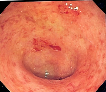
On a macroscopic basis, CMV Esophagitis may appear to be punched out lesions. Histology of CMV Esophagitis demonstrates enlarged cells with inclusions within both the cytoplasm and the nucleus of the cell.
The Patients Most at Risk
CMV esophagitis does not strike healthy individuals at random. It is a condition that emerges specifically in the context of a weakened immune system, where the body's natural defenses are too compromised to keep the cytomegalovirus in check. The vast majority of diagnosed patients carry an HIV diagnosis, making this infection one of the telling clinical markers of advanced immunosuppression. Beyond the HIV population, a significant share of cases arise among people whose immune systems have been deliberately or incidentally suppressed through other means.
Organ transplant recipients, who must take long-term immunosuppressive medication to prevent organ rejection, fall squarely into this vulnerable group. Patients managing diabetes or those on certain medications that blunt immune function also face elevated risk. The unifying thread across all these populations is clear: without a robust and responsive immune system, CMV can establish itself within the esophageal tissue and produce the painful, ulcerative disease that defines this condition.
How It Presents Clinically
The clinical picture of CMV esophagitis is dominated by the difficulty and pain of swallowing, a symptom known as dysphagia that often brings patients to medical attention. Upper abdominal pain is another hallmark, frequently worsening with each swallow and sometimes mimicking the burning sensation of heartburn. Nausea and vomiting round out the core symptom cluster, and in more severe presentations, patients may vomit blood—a condition called hematemesis that signals significant mucosal damage.
Diarrhea has also been reported as part of the broader picture. Fever may accompany the infection, reflecting the body's ongoing struggle against the viral invasion. Together, these symptoms paint a picture of an esophagus under active assault, where the lining is inflamed, ulcerated, and struggling to perform its basic function of safely moving food toward the stomach. The combination of painful swallowing, gastrointestinal distress, and occasional bleeding makes the condition both debilitating and, in its most acute forms, genuinely alarming for the patient.
Seeing the Damage: Diagnosis and Pathology
Confirming a diagnosis of CMV esophagitis relies heavily on endoscopy paired with tissue biopsy, and the findings are remarkably distinctive. When a physician looks down the esophagus during endoscopy, the hallmark appearance is that of large, punched-out lesions concentrated in the middle portion of the organ. These macroscopic ulcers are what set CMV esophagitis apart from other causes of esophageal inflammation. The story deepens under the microscope.
Histological examination reveals enlarged cells residing in the sub-epithelial layer, each harboring characteristic inclusions within both the nucleus and the cytoplasm. Aggregates of macrophage cells are also a common microscopic finding. To sharpen specificity further, fluorescent staining using an immunoperoxidase technique can be applied to the biopsy sample. Notably, radiologic imaging such as X-rays or CT scans does not help identify the infection itself, though it may reveal downstream complications like strictures or fistulas that the virus has provoked.
Fighting the Infection: Treatment and Its Toll
The therapeutic backbone for CMV esophagitis is intravenous ganciclovir, an antiviral agent delivered directly into the bloodstream to reach the infected esophageal tissue. When intravenous administration is not feasible, valganciclovir serves as an alternative, and foscarnet represents another option in the antiviral arsenal. However, the path to healing is not without its own physical cost. All three of these medications share a constellation of side effects that can compound the patient's existing misery: nausea, diarrhea, fever, and loss of appetite are common complaints.
In rarer but more concerning cases, patients may develop anemia or experience tremors, adding hematologic and neurological burdens on top of the original infection. The treatment landscape thus presents a difficult calculus—potent antivirals that can clear the esophageal ulcers but simultaneously tax the digestive system and, in some instances, the blood and nervous system. Managing these side effects becomes an integral part of the overall therapeutic strategy.
Did You Know?
- There are multiple ways cytomegalovirus can be transmitted. Mother to child transmission is common after childbirth.
- Transmission via saliva, tears, or skin contact is rare.
More in Inflammatory diseases of the digestive system
Sources
Compiled from Wikipedia and the sources listed below. Text from Wikipedia is available under CC BY-SA 4.0; this entry is adapted from it.
- Wikipedia: Cytomegalovirus esophagitis (CC BY-SA 4.0).
- Word definitions: the , each quoted from its Wikipedia article.
Spotted an error? Know more?
Reader corrections go straight into our review queue. Suggest an edit · How this site is sourced