Arnold–Chiari malformation
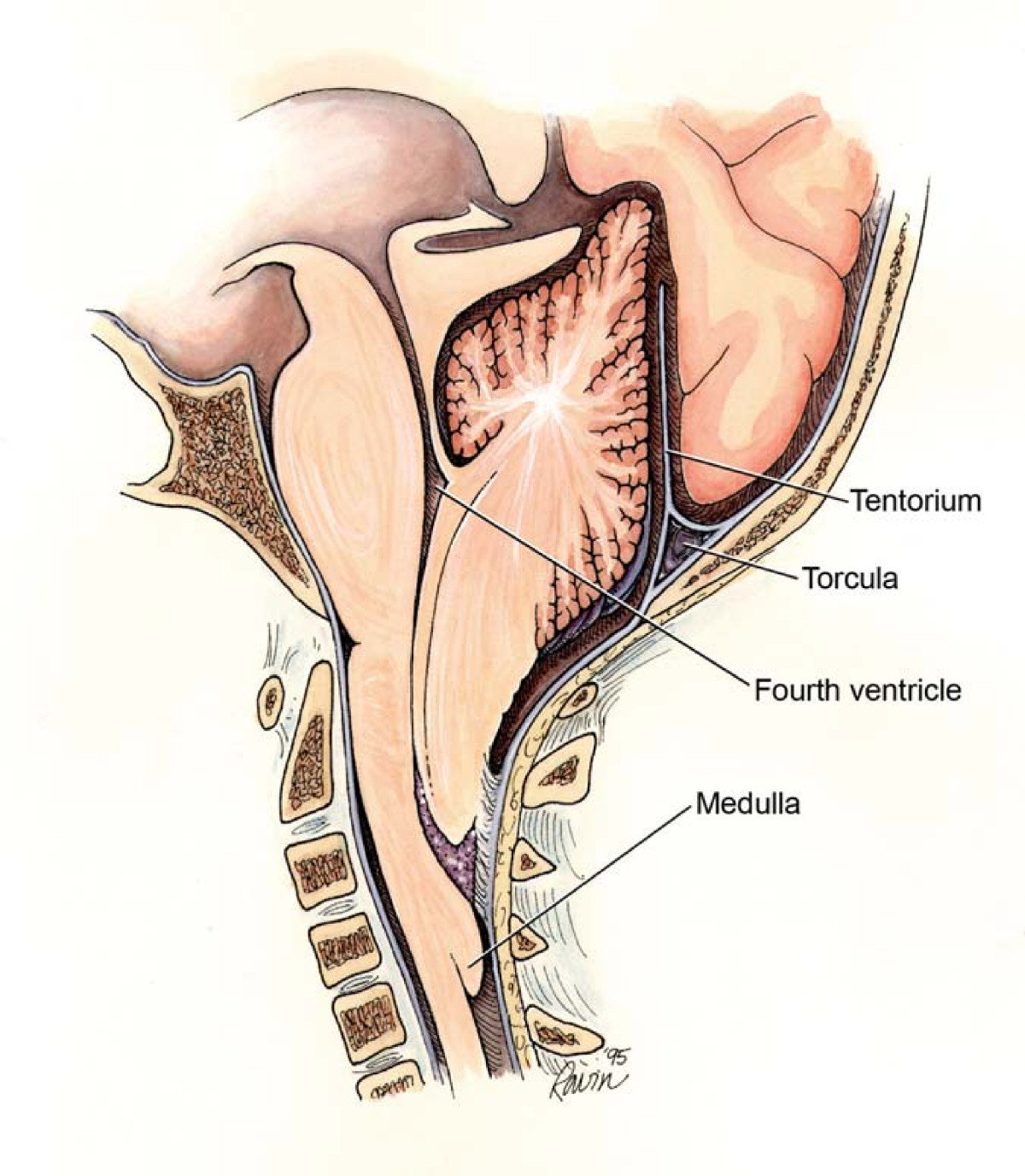
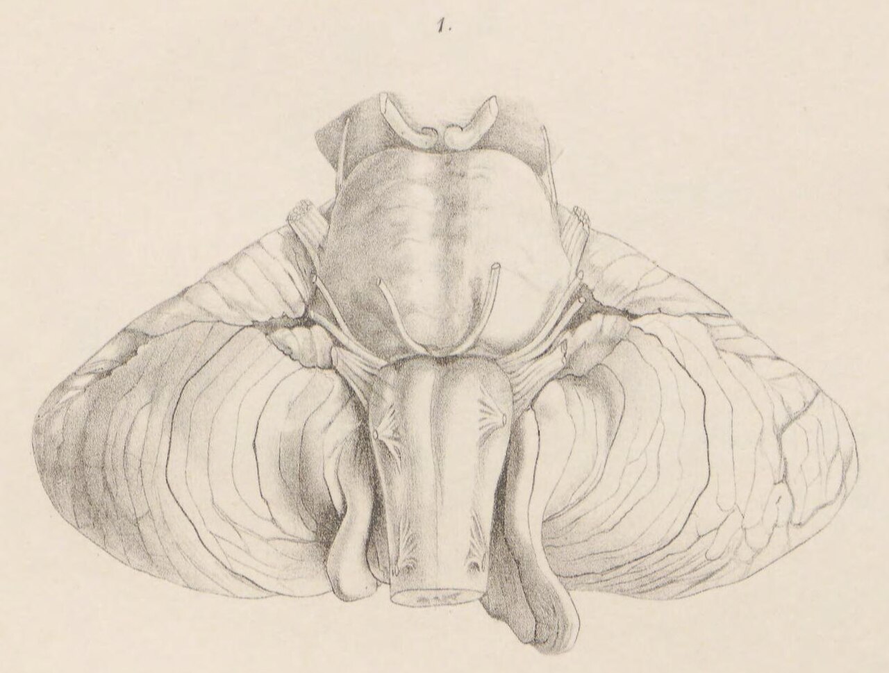
A type II Chiari malformation named after Chiari and Arnold.

see above · CC BY 2.0
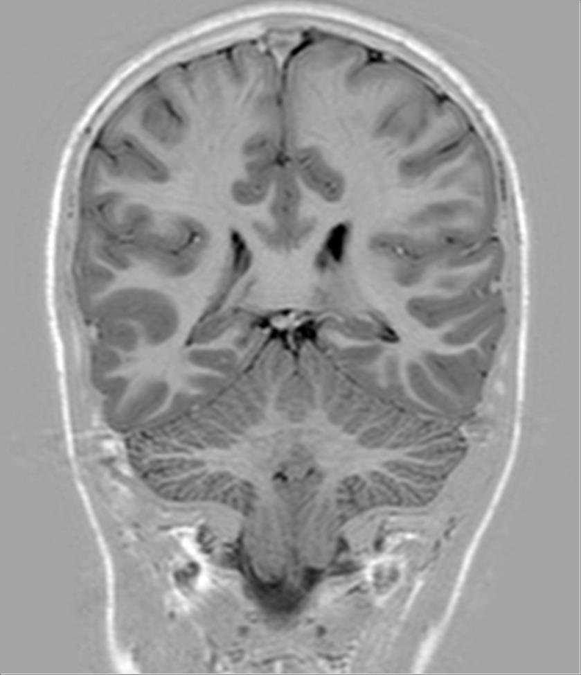
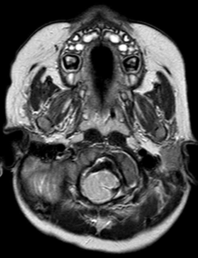
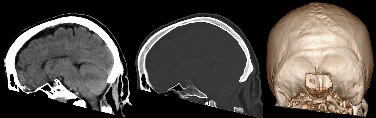
The Arnold–Chiari malformation is a term used to describe a type II Chiari malformation, a structural defect in the cerebellum characterized by downward displacement of cerebellar tonsils through the foramen magnum. It is named after the Austrian pathologist Hans Chiari and the German pathologist Julius Arnold, who contributed to its description. This condition can cause a range of neurological symptoms due to brainstem and lower cranial nerve dysfunction, including headaches, difficulty swallowing, and impaired coordination.
- field
- Neurology
- known_for
- Describing type II Chiari malformation (Arnold–Chiari malformation)
- related_conditions
- Chiari malformation, syringomyelia, hydrocephalus
Lore & Background
The malformation is named after the Austrian pathologist Hans Chiari and the German pathologist Julius Arnold. Chiari originally described seemingly related anomalies of the hindbrain in the late 19th century, including types I, II, and III. Later, other investigators added a fourth type. The Arnold–Chiari malformation specifically refers to type II, which involves both brainstem and tonsillar herniation through the foramen magnum.
Reader's Guide
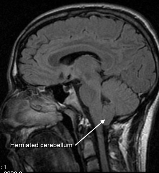
The Arnold–Chiari malformation is significant as a specific classification within Chiari malformations, representing a more severe form than type I. It is associated with symptoms such as headaches aggravated by Valsalva maneuvers, neurogenic dysphagia, and sleep apnea. Diagnosis is made through patient history, neurological examination, and medical imaging, with MRI being the preferred modality. The condition can lead to syringomyelia, a fluid-filled cyst in the spinal cord, present in 25% of type I patients. Understanding this malformation helps in managing symptoms and preventing complications like paralysis or hydrocephalus.
Did You Know?
- The Arnold–Chiari malformation is a type II Chiari malformation, named after Hans Chiari and Julius Arnold.
- Symptoms can include headaches aggravated by Valsalva maneuvers, difficulty swallowing, and sleep apnea.
- Syringomyelia is present in 25% of patients with type I Chiari malformations, which is related to type II.
- Diagnosis is made using MRI, which visualizes neural tissue and bone.
Anatomy, Naming & Classification
The Arnold–Chiari malformation is a structural abnormality rooted in the lower rear of the brain. At its core, the condition involves one or both cerebellar tonsils—the small, rounded extensions of the cerebellum—slipping downward through the foramen magnum, the bony opening at the skull's base where the brainstem meets the spinal cord. This downward displacement is what defines the malformation and sets the stage for every downstream neurological consequence. The condition carries the name of Austrian pathologist Hans Chiari, who first described the broader category of Chiari malformations. The specific type II variant, however, bears the additional name of German pathologist Julius Arnold, reflecting his parallel contributions to understanding this structural defect. Together, their nineteenth-century observations laid the groundwork for what is now recognized as a spectrum of congenital and acquired posterior fossa anomalies. The distinction between type I (tonsillar descent only) and type II (more extensive herniation) remains central to clinical classification and guides how physicians approach diagnosis and management.
Clinical Presentation & Symptom Spectrum
The neurological footprint of Chiari malformation is remarkably broad, reflecting the fact that the displaced tonsils compress the brainstem and lower cranial nerves. In type I, the hallmark complaint is a dull or throbbing occipital headache that characteristically worsens with Valsalva-type actions—coughing, sneezing, bearing down, or bending forward. Beyond pain, patients may report difficulty swallowing, unsteady gait, poor hand coordination, numbness or tingling in the extremities, speech difficulties, and dizziness. Less frequently, the condition manifests as tinnitus, cardiac rhythm disturbances, scoliosis linked to spinal cord impairment, or central sleep apnea. In the most severe presentations, pressure at the cervico-medullary junction can produce a progressive paralysis that spreads in a clockwise pattern, sequentially affecting the right arm, right leg, left leg, and left arm—or the reverse. Younger children tend to show a more rapid neurological decline with profound brainstem dysfunction over just days, whereas adults often present with subtler, more insidious symptoms. The variability in onset and expression makes early recognition particularly challenging.
Syringomyelia: The Spinal Cord Complication
A critical and often underappreciated complication of type I Chiari malformation is syringomyelia, a condition in which a fluid-filled cyst—called a syrinx—develops within the spinal cord. Approximately one in four patients with type I Chiari will develop this secondary lesion, most commonly between the C-4 and C-6 cervical levels, though the syrinx can extend upward into the medulla and pons or downward into the thoracic and lumbar segments. The prevailing theory holds that the herniated cerebellar tonsils create a mechanical plug at the foramen magnum, obstructing the normal outflow of cerebrospinal fluid from the brain into the spinal canal. This trapped fluid pressure then drives the formation of the cyst. Symptoms of syringomyelia include pain, weakness, numbness, and stiffness in the back, shoulders, arms, or legs, along with loss of temperature sensation, abnormal sweating, sexual dysfunction, and impaired bowel or bladder control. While the condition is typically chronic and progressive, some individuals experience a stable syrinx that causes minimal or no symptoms throughout their entire lives.
Pathophysiology: Three Mechanisms of Tonsillar Descent
The mechanisms behind Chiari malformation are not singular but span at least three distinct pathophysiological pathways. The most frequent cause in type I is a congenitally undersized posterior fossa, the bony compartment at the skull's base that houses the cerebellum. A second pathway involves elevated pressure above the foramen magnum pushing the cerebellum downward; contributors include hydrocephalus, brain tumors, subdural hematomas, arachnoid cysts, craniosynostosis (premature fusion of cranial sutures, particularly the lambdoid suture), and hyperostotic bone-growth disorders such as osteopetrosis. A third mechanism operates in the opposite direction: negative pressure or a pulling force from below the foramen magnum draws the tonsils downward. This can result from a tethered spinal cord, an excessively tense filum terminale, a cerebrospinal fluid leak creating subarachnoid negative pressure, or a CSF-venous fistula. Additionally, traumatic brain injury has been linked to delayed acquired Chiari malformation, though the precise mechanism remains unclear, and the condition may stay asymptomatic for years before a whiplash or other event triggers symptom onset.
Gallery
Frequently Asked Questions
Who is Arnold–Chiari malformation?
It is a congenital structural abnormality in which the cerebellar tonsils herniate below the foramen magnum, and it bears the names of two 19th-century pathologists—German physician Julius Arnold and Austrian pathologist Hans Chiari—who each contributed to describing its anatomical features.
What is Arnold–Chiari malformation's role in the body?
Rather than serving a physiological purpose, it acts as a mechanical obstruction: the displaced cerebellar tissue compresses the brainstem and lower cranial nerves, disrupting normal neurological signaling. It is classified as a type II Chiari malformation and frequently co-occurs with spina bifida in affected individuals.
What 'powers' does Arnold–Chiari malformation wield?
Its neurological effects include chronic headaches, impaired coordination and balance, and difficulty swallowing due to lower cranial nerve compression. It is also strongly linked to the development of syringomyelia and hydrocephalus, which compound the patient's overall symptom burden.
How does Arnold–Chiari malformation's story end?
There is no single narrative arc; outcomes range from posterior-fossa surgical decompression to manage symptoms, to lifelong neurological monitoring when the malformation is mild. Without intervention, progressive brainstem compression can lead to serious and potentially life-threatening neurological decline.
Why is Arnold–Chiari malformation important in the Neurology 'universe'?
It remains a cornerstone diagnosis in pediatric and adult neurosurgery because it links posterior-fossa anatomy to a cascade of neurological deficits. Understanding it helped establish the relationship between cerebellar positioning, cerebrospinal-fluid dynamics, and conditions like syringomyelia.
More in Diseases named after discoverers 1-24
Spotted an error? Know more?
This is a living reference — every entry is fact-audited, and reader corrections feed straight into our audit queue. Suggest an edit · See this site's audit record