Alexander disease
Rare leukodystrophy causing white matter destruction and Rosenthal fibers.

Wikimedia Commons · CC BY 4.0
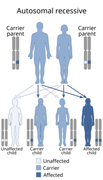
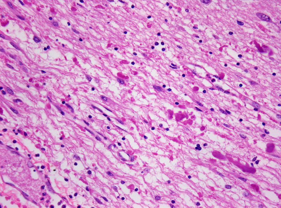
Alexander disease is an extremely rare genetic disorder that falls under the category of leukodystrophies—conditions that damage the myelin sheath protecting nerve fibers in the brain. It is inherited in an autosomal dominant pattern, meaning a child of an affected parent has a 50% chance of developing the disease if the parent carries one copy of the mutated gene, though most cases arise from new, spontaneous mutations. The hallmark of the disease is the breakdown of white matter in the brain, along with the buildup of abnormal protein clumps called Rosenthal fibers inside astrocytes.
The condition was first described in 1949 by Dr. W. Stewart Alexander, who treated a 15-month-old infant with an enlarged head, fluid on the brain, seizures, and developmental delays. Between 1949 and 1964, fifteen more similar cases were reported, leading researchers to recognize them as the same disease and name it after Alexander. It affects both males and females equally, with no differences based on ethnicity, race, geography, or socioeconomic background. Alexander disease is progressive and often fatal.
Symptoms vary widely. The most common form appears in infancy, usually within the first two years of life, and includes delays in physical and mental development, loss of previously gained milestones, abnormal head enlargement, and seizures. The juvenile form begins between ages 2 and 13 and may involve excessive vomiting, trouble swallowing and speaking, poor coordination, and loss of motor control. Adult-onset forms are rarer and can mimic Parkinson’s disease, multiple sclerosis, or appear primarily as a psychiatric disorder.
In early-onset or neonatal cases, symptoms include seizures, fluid buildup in the brain, high protein levels in cerebrospinal fluid, and severe motor and intellectual impairment. In type I Alexander disease (onset before age 4), symptoms include seizures, enlarged brain and head, limb stiffness, delayed intellectual and physical development, recurrent vomiting, and difficulty gaining weight. In type II Alexander disease (onset after age 4), symptoms include speech problems, trouble swallowing, poor coordination, scoliosis, recurrent vomiting, and difficulty gaining weight.
The disease is caused by a gain-of-function mutation in the GFAP gene, located on chromosome 17q21. This mutation leads to protein aggregates called Rosenthal fibers forming.
- first_documented
- 1949
- first_documented_by
- W. Stewart Alexander
- inheritance
- Autosomal dominant
- prevalence
- 1 in 2.7 million
- types
- Infantile, juvenile, adult; also Type I and Type II
- gene
- GFAP on chromosome 17q21
Lore & Background
Alexander disease was first identified in 1949 by W. Stewart Alexander, who treated a 15-month-old infant with megalencephaly, hydrocephaly, seizures, and developmental delays. Between 1949 and 1964, 15 more similar cases were observed, leading to the suggestion that these cases represented the same disease and that it be named after Alexander. The disease occurs in both males and females, with no ethnic, racial, geographic, or cultural/economic differences in its distribution.
Reader's Guide
Alexander disease is a progressive and often fatal leukodystrophy caused by mutations in the GFAP gene, inherited in an autosomal dominant manner, though most cases arise de novo. It is classified by age at onset into infantile, juvenile, and adult forms, or alternatively into Type I (onset before age 4) and Type II (onset after age 4). Symptoms vary widely but include developmental delays, seizures, macrocephaly, spasticity, and in adult forms, symptoms mimicking Parkinson's disease, multiple sclerosis, or psychiatric disorders. Diagnosis is made via MRI, genetic testing, or clinical symptoms. Prognosis is poor: infantile form patients usually die before age seven, with an average duration of about three years; juvenile form duration is about six years. Zilganersen (Zanvastro) was approved for medical use in the United States in September 2026. The disease is very rare, with an estimated prevalence of 1 in 2.7 million.
Did You Know?
- Alexander disease was first documented in 1949 by W. Stewart Alexander, who treated a 15-month-old infant.
- The disease is caused by mutations in the GFAP gene on chromosome 17q21, inherited in an autosomal dominant manner.
- Rosenthal fibers—abnormal clumps of protein in astrocytes—are a hallmark of Alexander disease.
- Zilganersen (Zanvastro) was approved for medical use in the United States in September 2026.
Clinical Presentation Across the Lifespan
Alexander disease manifests differently depending on when it first appears, and its symptoms span a wide spectrum of neurological and developmental disruption. In its most common infantile presentation, the condition typically emerges within the first two years of life, bringing with it delays in both mental and physical growth, a troubling regression of previously achieved milestones, an abnormal enlargement of the head, and recurrent seizures. The juvenile form, which surfaces between the second and thirteenth years, tends to produce excessive vomiting, struggles with swallowing and speech, deteriorating coordination, and a gradual loss of voluntary motor control. Adult-onset cases present a particularly diagnostic challenge because their symptoms can closely resemble those of Parkinson's disease, multiple sclerosis, or even a primary psychiatric condition. Researchers have further subdivided the disease into Type I, appearing before age four with limb stiffness, an enlarged brain, and difficulty gaining weight, and Type II, emerging after age four with scoliosis, speech impairment, and persistent vomiting. Some scholars have also proposed a neonatal category for cases whose symptoms were already evident before or at birth, marked by fluid accumulation in the brain and severe intellectual and motor impairment.
Genetic Origins and Cellular Pathology
At its molecular core, Alexander disease is driven by a gain-of-function mutation in the gene encoding glial fibrillary acidic protein, located on chromosome 17q21. The condition follows an autosomal dominant inheritance pattern, meaning a child of a heterozygous affected parent carries a fifty percent chance of inheriting the faulty gene. In practice, however, the majority of diagnosed cases stem from sporadic, de novo mutations rather than familial transmission. The mutated GFAP protein triggers the accumulation of dense, eosinophilic protein clumps called Rosenthal fibers within the cytoplasm of astrocytes, though the precise biochemical pathway behind their formation remains incompletely understood. These fibers are not exclusive to Alexander disease—they also appear in certain cancers, Alzheimer's, Parkinson's, Huntington's, and ALS—but their spatial distribution and concentration in Alexander disease are distinct from those seen in any other condition. The disease primarily targets the midbrain and cerebellum, and the progressive destruction of white matter is compounded by an overload of long-chain fatty acids that further dismantle the myelin sheath insulating nerve fibers.
The Diagnostic Puzzle
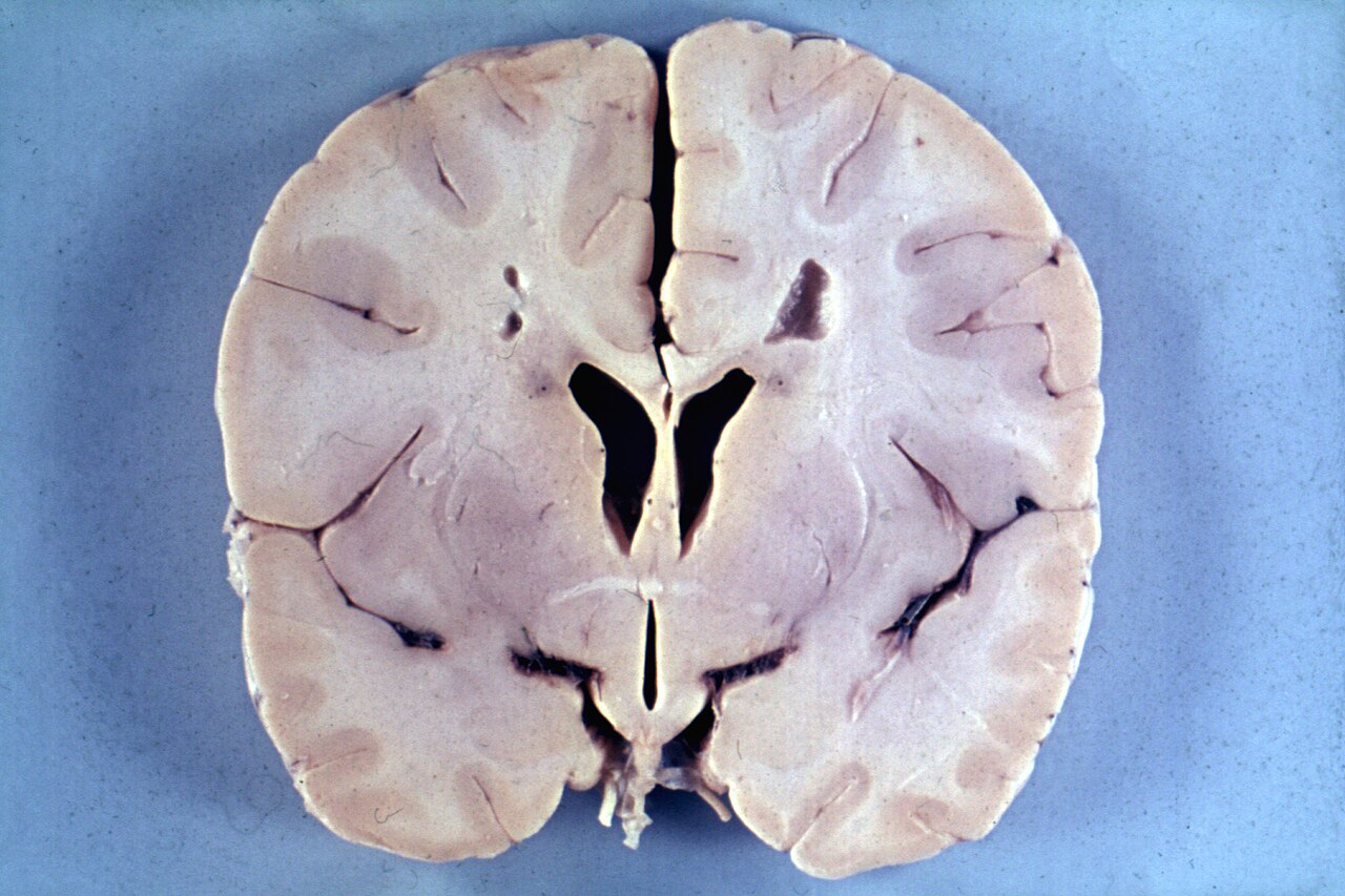
Confirming a diagnosis of Alexander disease can be an arduous process, particularly in adult patients whose symptoms overlap with far more common neurological disorders. Magnetic resonance imaging serves as the principal imaging tool, revealing characteristic patterns such as periventricular white matter changes, atrophy of the medulla, and signal abnormalities extending into the spinal cord. Computed tomography scans may additionally show reduced white matter density concentrated in the frontal lobes and dilation of the lateral ventricles. Genetic testing for the underlying GFAP mutation offers a definitive molecular confirmation, while a working clinical diagnosis can sometimes be assembled from the constellation of an enlarged head, progressive neurological decline, and the exclusion of other leukodystrophies through negative laboratory results. Despite these tools, many adults endure years of misdiagnosis—being labeled with multiple sclerosis, Parkinson's disease, or a psychiatric condition—before an MRI finally uncovers the distinctive white matter pathology. The rarity of the disease, affecting roughly one in twenty-seven million individuals, compounds the difficulty, as many clinicians may never encounter a confirmed case in their entire careers.
Prognosis, Rarity, and the Road to Treatment
The outlook for individuals living with Alexander disease remains sobering. Those affected by the infantile form typically succumb before reaching their seventh birthday, with an average survival of roughly three years from onset. The juvenile form carries a somewhat longer trajectory, averaging around six years. A consistent pattern emerges across all presentations: the later the disease manifests, the more gradual its progression. The condition shows no bias by sex, ethnicity, race, geography, or socioeconomic background, striking with equal indifference across all populations. Its extreme rarity—an estimated prevalence of one in 2.7 million—means that even among the many forms of leukodystrophy, it occupies a particularly obscure corner of neurological medicine. Historically, the disease was first described in 1949 by W. Stewart Alexander, who treated a fifteen-month-old infant with an enlarged head, seizures, and developmental delays; over the next fifteen years, only fifteen additional similar cases were recorded before the condition was formally named in his honor. A glimmer of therapeutic hope arrived in September 2026, when the United States approved zilganersen, marketed as Zanvastro, for medical use, marking the first targeted treatment for this devastating disorder.
Gallery


Frequently Asked Questions
Who is Alexander disease?
Alexander disease is a rare genetic leukodystrophy first formally described in 1949 by neurologist W. Stewart Alexander, who identified the characteristic white matter degeneration and Rosenthal fiber accumulation in his patients. The condition bears his name as the physician who first documented its distinct clinical and pathological profile.
What are Alexander disease's powers/role?
The disorder progressively destroys the brain's white matter by damaging the myelin sheath that insulates nerve fibers, while simultaneously causing astrocytes to accumulate abnormal protein clumps called Rosenthal fibers. It expresses itself in three recognized forms—infantile, juvenile, and adult—each carrying a different severity and tempo of neurological decline.
How does Alexander disease's story end?
The infantile form is the most aggressive, often producing severe motor and cognitive deterioration within the first few years of life, whereas the adult variant may progress very gradually over decades. No curative therapy currently exists, so management centers on controlling seizures, spasticity, and other downstream symptoms.
Why is Alexander disease important?
At roughly one case per 2.7 million individuals, it ranks among the rarest leukodystrophies, yet it offers a unique window into GFAP protein biology and astrocyte maintenance. Research into the condition has deepened understanding of how a single gene mutation on chromosome 17q21 can unravel normal white matter integrity.
What's Alexander disease's origin story?
The disease follows an autosomal dominant inheritance pattern, so a single mutated copy of the GFAP gene is sufficient to trigger it, and a child of an affected parent faces a 50 percent chance of inheriting the variant. In practice, however, the majority of cases arise from spontaneous de novo mutations rather than being passed down through a family line.
More in Diseases named after discoverers 1-24
Spotted an error? Know more?
This is a living reference — every entry is fact-audited, and reader corrections feed straight into our audit queue. Suggest an edit · See this site's audit record