Adams–Oliver syndrome
Rare congenital disorder with scalp, limb, and skin defects.

Adams–Oliver syndrome (AOS) is a rare condition present from birth. Its main features include missing skin on the scalp (sometimes with an underlying skull defect), shortened or missing parts of the limbs, and a blotchy, mottled appearance of the skin.
The scalp defect, known as aplasia cutis congenita, can range from a small bald patch to a large area where the brain is exposed. Limb defects vary as well, from shortened fingers or toes to more severe reductions in the arms or legs. People with AOS may be shorter than average, though still within the low-normal range. The skin often has a marbled look (cutis marmorata telangiectatica congenita). Other problems can occur, including heart defects, cleft lip or palate, kidney abnormalities, and neurological issues such as seizures or developmental delays. Blood vessel abnormalities have also been described, affecting arteries like the aortic arch, middle cerebral, and pulmonary arteries, as well as veins such as the portal vein and umbilical veins.
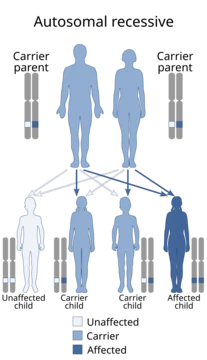
Genetically, AOS can be inherited in different ways. In some families, it follows an autosomal dominant pattern, meaning one copy of a mutated gene can cause the condition. In other families, it appears to be autosomal recessive, requiring two mutated copies. The severity can vary widely even within the same family. Six genes have been linked to AOS: ARHGAP31, DOCK6, RBPJ, EOGT, NOTCH1, and DLL4. ARHGAP31 and DOCK6 are involved in regulating the Rho family of GTPases, which control cell shape and movement. Dominant mutations in ARHGAP31 and recessive mutations in DOCK6 lead to problems with the cytoskeleton. The other four genes—RBPJ, EOGT, NOTCH1, and DLL4—are part of the Notch signaling pathway, which is important for cell development. Mutations in EOGT are recessive, while mutations in the other three are dominant.
The exact cause of the birth defects in AOS is not known. Because similar limb and heart problems occur in animal models when the fetus is deprived of oxygen early in pregnancy, and because blood vessel issues are common in AOS, researchers think the condition may result from a problem with blood vessel formation (vasculogenesis). In rare cases, AOS has been linked to chromosomal translocations. Tests on other candidate genes (such as ALX4, ALX1, MSX1, MSX2, P63, RUNX2, and HOXD13) did not find disease-causing mutations. The discovery of mutations in
- field
- Medical genetics
- known_for
- First description of Adams–Oliver syndrome
- reported_by
- Forrest H. Adams and Clarence Paul Oliver
- inheritance
- Autosomal dominant and autosomal recessive
- estimated_prevalence
- Approximately 1 in 225,000 live births
Lore & Background
Adams–Oliver syndrome (AOS) was first reported by the American pediatric cardiologist Forrest H. Adams and the clinical geneticist Clarence Paul Oliver in a family with eight affected members. The condition is characterized by aplasia cutis congenita (missing skin, typically at the vertex of the skull) and terminal transverse limb defects, such as shortened digits. Additional features may include mottled skin, mild growth deficiency, and various congenital anomalies involving the cardiovascular system, cleft lip or palate, renal abnormalities, and neurological disorders.
Reader's Guide
Adams–Oliver syndrome is significant as a rare genetic disorder that highlights the roles of the Rho family of GTPases and the Notch signaling pathway in human development. Six genes have been identified: ARHGAP31, DOCK6, RBPJ, EOGT, NOTCH1, and DLL4. Mutations in these genes disrupt cytoskeletal regulation or Notch signaling, leading to the characteristic defects. The diagnosis is clinical, based on major and minor criteria, with genetic testing accounting for only about 50% of cases. Management is symptomatic, and prognosis is generally excellent, though severe scalp defects can lead to complications. The syndrome underscores the complexity of genetic inheritance, with both autosomal dominant and recessive forms described.
Did You Know?
- Adams–Oliver syndrome was first reported by Forrest H. Adams and Clarence Paul Oliver in a family with eight affected members.
- The condition involves aplasia cutis congenita, typically at the vertex of the skull, and terminal transverse limb defects.
- Six genes have been identified: ARHGAP31, DOCK6, RBPJ, EOGT, NOTCH1, and DLL4.
- Genetic testing identifies mutations in only about 50% of patients with AOS.
The Clinical Portrait
Adams–Oliver syndrome presents as a constellation of congenital abnormalities that, taken together, form a recognizable clinical picture. At the core of the condition lie two hallmark features: aplasia cutis congenita, in which skin is absent at the vertex of the skull, and terminal transverse limb defects that can shorten or truncate digits of the hands, feet, or both. The scalp lesions themselves show remarkable variability, ranging from small, solitary, round patches of missing hair to the extreme scenario of fully exposed cranial contents. Beyond these defining traits, affected individuals often display a mottled, net-like skin pattern known as cutis marmorata telangiectatica congenita, along with mild growth deficiency placing height in the low-normal range. A broader spectrum of associated anomalies has been documented, including cardiovascular malformations, cleft lip or palate, renal abnormalities, and neurologic issues such as seizure disorders and developmental delay. Vascular involvement is particularly notable, with reports of a hypoplastic aortic arch, absent portal vein, arteriovenous malformations, and dilated renal veins, underscoring the syndrome's deep connection to the circulatory system.
Genetic Architecture & Inheritance
The inheritance pattern of Adams–Oliver syndrome was initially understood as autosomal dominant, based on observations of multiple affected individuals spanning several generations within the same family. However, researchers noted that severity varied considerably among relatives, pointing to variable expressivity and reduced penetrance of the causative allele. Later evidence revealed that a subset of cases follows an autosomal recessive pattern, often presenting with somewhat more severe phenotypic effects. To date, six genes have been linked to the condition: ARHGAP31, DOCK6, RBPJ, EOGT, NOTCH1, and DLL4. ARHGAP31 and DOCK6 function as regulatory proteins governing members of the Rho family of GTPases, specifically modulating Cdc42 and Rac1 activity. Mutations in ARHGAP31, a GTPase-activating protein, follow a dominant pattern, while DOCK6, a guanine nucleotide exchange factor, shows recessive inheritance; both ultimately cause an accumulation of inactive GTPase that disrupts cytoskeletal integrity. The remaining four genes—RBPJ, EOGT, NOTCH1, and DLL4—operate within the Notch signalling pathway, with EOGT mutations showing recessive inheritance and the other three associated with dominant inheritance.
The Vascular Hypothesis & Pathogenic Mechanisms
The exact molecular mechanism producing the congenital abnormalities seen in Adams–Oliver syndrome remains unresolved. One compelling line of reasoning draws on animal models in which hypoxic insults during the first trimester generate terminal transverse limb anomalies and cardiovascular malformations strikingly similar to those observed in AOS patients. Coupled with the frequent co-occurrence of cardiac and vascular defects in affected individuals, researchers have proposed that the full spectrum of AOS manifestations may stem from a fundamental disorder of vasculogenesis, the process by which blood vessels form during embryonic development. Early attempts to identify causative mutations in a panel of candidate genes, including ALX4, ALX1, MSX1, MSX2, P63, RUNX2, and HOXD13, proved unfruitful. In rare instances, chromosomal translocations have been associated with the syndrome. The breakthrough came with the identification of six disease-associated genes, which collectively spotlight the Rho family of GTPases and the Notch signalling pathway as central players in the pathogenesis of this condition, shifting the field's understanding from a vague structural defect to a more defined molecular framework.
Diagnosis, Care & Long-Term Outlook
Because Adams–Oliver syndrome is a clinical diagnosis, physicians rely on a structured system of major and minor criteria rather than a single definitive test. The presence of two major features is considered sufficient to confirm the diagnosis, while one major combined with one minor feature raises strong suspicion. Genetic testing can be performed to detect mutations in the six known AOS genes, yet these account for only an estimated half of all affected patients, meaning a definitive molecular diagnosis may remain elusive in many cases. Treatment is predominantly symptomatic, tailored to the specific congenital anomalies each individual carries. When scalp or cranial bone defects are severe, early surgical intervention with grafting becomes necessary to protect the underlying brain. Despite the complexity of the condition, the overall prognosis is encouraging: most children with Adams–Oliver syndrome can be expected to live a normal lifespan. The principal risk arises in those with the most severe cranial involvement, where complications such as hemorrhage or meningitis can lead to long-term disability. The syndrome remains exceedingly rare, with only 100 to 200 cases documented in the medical literature and an estimated incidence of roughly one in 225,000 live births.
Frequently Asked Questions
Who is Adams–Oliver syndrome?
It is a rare congenital condition that falls under medical genetics, first formally described by physicians Forrest H. Adams and Clarence Paul Oliver. It can be passed down through either autosomal dominant or autosomal recessive inheritance patterns.
What are Adams–Oliver syndrome's signature traits?
Its hallmark features include aplasia cutis congenita (absent scalp skin, sometimes with an underlying skull defect), limb reductions that range from shortened digits to more severe arm or leg involvement, and a distinctive mottled, marbled skin appearance. Affected individuals are often shorter than average, though still within the low-normal range.
How common is Adams–Oliver syndrome in the broader 'universe'?
It is estimated to affect roughly 1 in 225,000 live births, making it an exceedingly rare entry in the catalog of named genetic conditions.
What is Adams–Oliver syndrome's origin story?
The syndrome takes its name from the two physicians who first documented the constellation of scalp, limb, and skin findings in affected newborns, establishing it as a recognizable entity in medical genetics.
How does Adams–Oliver syndrome's 'story' play out for those affected?
Severity varies considerably from person to person: the scalp defect can be a small bald patch or a large area with exposed brain tissue, and limb involvement may be limited to shortened fingers or extend to more substantial reductions. Despite these challenges, many individuals live into adulthood with appropriate medical support.
More in Diseases named after discoverers 1-24
Spotted an error? Know more?
This is a living reference — every entry is fact-audited, and reader corrections feed straight into our audit queue. Suggest an edit · See this site's audit record