
Brain
Central organ of the nervous system in vertebrates and most invertebrates.

Bobjgalindo · CC BY-SA 4.0
The brain is the central command center of the nervous system, found in all vertebrates and most invertebrates. Made of nervous tissue, it sits in the head, close to the sense organs for sight, hearing, and smell. As the body’s most specialized organ, it takes in sensory information, processes it into thought, cognition, and intelligence, and coordinates muscle movements and hormone release via the endocrine system.
Invertebrate brains are built from paired segmental ganglia along the ventral nerve cord, each ganglion handling only its own body segment. Vertebrate brains, in contrast, develop from the dorsal nerve cord as a swollen vesicle at the front end of the neural tube, giving them centralized control over all body segments. Every vertebrate brain can be split into three embryonic parts: the forebrain (prosencephalon, which further divides into telencephalon and diencephalon), the midbrain (mesencephalon), and the hindbrain (rhombencephalon, split into metencephalon and myelencephalon). The spinal cord, which manages basic body functions below the head, is essentially a tail-end extension of the myelencephalon, housed inside the backbone. Together, the brain and spinal cord form the central nervous system in all vertebrates.
In humans, the cerebral cortex holds about 14–16 billion neurons, while the cerebellum contains an estimated 55–70 billion. Each neuron connects to thousands of others via synapses, communicating through thread-like extensions called dendrites and axons. Axons are usually wrapped in myelin and carry rapid electrical pulses—action potentials—to specific target cells elsewhere in the brain or body. The prefrontal cortex, which handles executive functions, is especially developed in humans.
Physiologically, the brain exerts centralized control over the body’s other organs. It directs muscle activity patterns and drives hormone secretion, enabling quick, coordinated responses to environmental changes. Simple reflexes can be handled by the spinal cord or peripheral ganglia, but complex, purposeful behavior based on sensory input requires the brain’s ability to integrate information.
The workings of individual brain cells are now well understood, but how millions of them cooperate remains unsolved. Modern neuroscience often models the brain as a biological computer—mechanically very different from a digital one, but similar in that it acquires, stores, and processes information from the world.
This article compares brains across the animal kingdom, focusing on vertebrates. It covers the human brain only where it shares features with other brains; differences, along with brain disease and damage effects, are addressed in the human brain article.
**Structure**
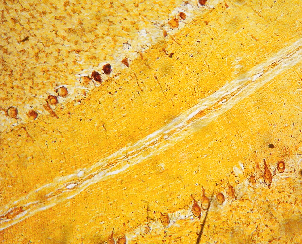
Brain shape and size vary greatly between species, making common features hard to identify. Still, several architectural principles apply widely. Some structural aspects are nearly universal; others separate advanced brains from simpler ones, or vertebrates from invertebrates. Basic anatomy can be seen by eye, but more advanced techniques exist. Fresh brain tissue is too soft to work with, so it’s hardened in alcohol or other fixatives, then sliced to reveal the interior. Inside, darker grey matter areas are separated by lighter white matter. Staining slices with chemicals highlights regions rich in specific molecules. Microscopy reveals microstructure, and tracing techniques map connections between brain areas.
**Cellular structure**
All brains contain two main cell types: neurons and glial cells. Glia come in several forms and provide structural support, metabolic help, insulation, and developmental guidance. Neurons are typically considered the most important. In humans, the cerebral cortex has about 14–16 billion neurons, and the cerebellum 55–70 billion. Each neuron connects to thousands of others via synapses. Their unique ability is sending signals to specific targets, sometimes over long distances, through a thin protoplasmic fiber called an axon. The axon extends from the cell body, branching to nearby or distant brain regions or body parts. An axon can be remarkably long—for instance, if a pyramidal cell of the cerebral cortex were magnified...
- type
- Organ
- location
- Head (cephalization)
- constituent_cells
- Neurons and glial cells
- human_cortical_neurons
- 14–16 billion
- human_cerebellar_neurons
- 55–70 billion
Lore & Background
The brain arises differently in vertebrates and invertebrates. Invertebrate brains develop from paired segmental ganglia of the ventral nerve cord, each responsible for a body segment. Vertebrate brains develop axially from the midline dorsal nerve cord as a vesicular enlargement at the rostral end of the neural tube, with centralized control over all body segments. All vertebrate brains can be embryonically divided into three parts: forebrain, midbrain, and hindbrain. The spinal cord, which coordinates somatic functions below the head, is a caudal extension of the myelencephalon enclosed in the vertebral column; together, the brain and spinal cord form the central nervous system in all vertebrates.
Reader's Guide
The brain exerts centralized control over a body's other organs by generating patterns of muscle activity and driving hormone secretion, allowing rapid and coordinated responses to environmental changes. While reflexes can be mediated by the spinal cord or peripheral ganglia, sophisticated purposeful behavior requires the information-integrating capabilities of a centralized brain. The operations of individual brain cells are understood in considerable detail, but how they cooperate in ensembles of millions remains unsolved. Modern neuroscience treats the brain as a biological computer, different in mechanism from a digital computer but similar in acquiring, storing, and processing information. The human brain shares properties with other brains; topics such as brain disease and effects of brain damage are covered in the human brain article.
Did You Know?
- The human cerebral cortex contains approximately 14–16 billion neurons, and the cerebellum contains 55–70 billion.
- Each neuron is connected by synapses to several thousand other neurons.
Architecture of the Central Command
The human brain is the master control organ of the nervous system, paired with the spinal cord to form the central nervous system. At roughly 1.2 to 1.4 kilograms—about two percent of body weight—it is a remarkably soft, gel-like structure enclosed by the skull and wrapped in three meningeal membranes: the dura mater, arachnoid mater, and pia mater. Its architecture splits into three principal divisions: the cerebrum, the brainstem, and the cerebellum. The cerebrum is by far the largest, built from two hemispheres, each with a white-matter core wrapped in a grey-matter cortex. That cortex layers into a six-layered neocortex and a thinner three-or-four-layer allocortex. Each hemisphere is divided into frontal, parietal, temporal, and occipital lobes; the frontal lobe handles executive functions such as planning and self-control, while the occipital lobe is dedicated to vision. Although the hemispheres are broadly symmetrical, some functions are lateralized—language typically on the left, visual-spatial processing on the right. They are linked by commissural tracts, the largest being the corpus callosum. The brainstem, made up of the midbrain, pons, and medulla oblongata, connects the cerebrum to the spinal cord, and the cerebellum sits behind, joined by three pairs of peduncles. Deeper still lie paired structures like the thalamus, hypothalamus, amygdalae, and hippocampi.
The Cellular Engine
Inside this soft architecture lives an astonishing cellular population. The brain contains more than 86 billion neurons, accompanied by a roughly equal number of supportive glial cells. These neurons do not work in isolation; they interconnect to form neural pathways, neural circuits, and elaborate network systems that span the entire organ. The fundamental process driving all of this connectivity is neurotransmission: when a nerve impulse reaches a neuron, it triggers the release of neurotransmitters into the synaptic gap, passing the signal onward to the next cell. This electrochemical relay is what makes every thought, movement, and sensation possible. The sheer scale of the interconnections means that even a small region of cortex can participate in vast, distributed networks. The ventricular system—four interconnected chambers within the cerebrum—produces and circulates cerebrospinal fluid, which also provides mechanical cushioning. The blood–brain barrier, maintained in part by the glia limitans, the basement membrane of the pia mater, isolates the brain's delicate chemistry from the general bloodstream, ensuring that the precise concentration of neurotransmitters and other molecules needed for signaling is preserved. This barrier is one of the brain's most critical protective mechanisms, yet it also presents a challenge for delivering therapeutic drugs to the brain.
Vulnerability and the Spectrum of Disease
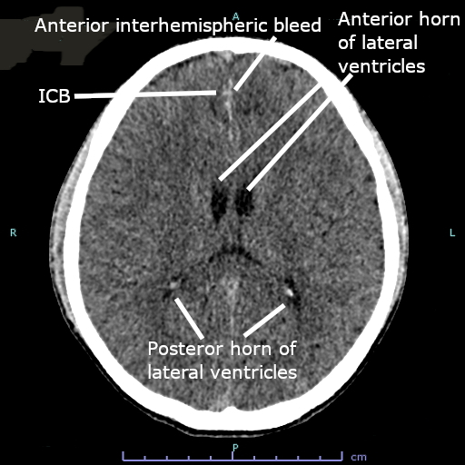
Despite the skull, the cerebrospinal fluid cushion, and the blood–brain barrier, the brain remains remarkably vulnerable. Traumatic injury and stroke—a sudden loss of blood supply to brain tissue—can cause devastating damage. Beyond acute events, the brain is prone to a range of degenerative disorders, including Parkinson's disease, multiple sclerosis, and the dementias such as Alzheimer's disease, which progressively erode cognitive function. Psychiatric conditions like schizophrenia and clinical depression are also thought to arise from underlying brain dysfunctions, though their exact mechanisms remain complex. Tumors can also develop within the brain, both benign and malignant, though most originate from sites elsewhere in the body before metastasizing. The brain's susceptibility is a direct consequence of its extraordinary metabolic demands and the fragility of its cellular networks. A single disrupted pathway can cascade into widespread dysfunction, which is why even localized damage can produce effects far beyond the injured region. Understanding these vulnerabilities has driven much of modern neuroscience, as researchers work to identify the specific circuits and molecular processes that break down in each condition.
Studying the Mind's Hardware
The scientific study of the brain splits into two complementary disciplines: neuroanatomy, which maps its physical structure, and neuroscience, which investigates how it functions. For centuries, much of what we know came from examining specimens of other animals under the microscope. In the modern era, functional neuroimaging and electroencephalography recordings have opened windows into the living brain, allowing researchers to observe activity in real time. The medical histories of patients who suffered brain injuries have also been invaluable, revealing which functions are tied to which regions. Beyond the laboratory, the brain has long captivated the human imagination. The philosophy of mind has grappled for centuries with the nature of consciousness and the mind–body problem. In the nineteenth century, the pseudoscience of phrenology attempted to map personality traits onto specific cortical regions. These cultural echoes remind us that the brain is not merely a biological organ but a central symbol of what it means to be human.
Gallery
Frequently Asked Questions
What is the brain in animal anatomy?
The brain is the central organ of the nervous system found in all vertebrates and most invertebrate species. It is composed of nervous tissue and represents the most specialized organ in the animal body.
Where is the brain located in animals?
In nearly all animals, the brain sits in the head region, a pattern known as cephalization. This positioning places it close to key sensory organs for vision, hearing, and smell, allowing rapid access to incoming environmental data.
What types of cells make up the brain?
The brain is built from two main cell types: neurons, which transmit and process electrical signals, and glial cells, which provide structural and metabolic support. Together these cells form the functional tissue of the entire organ.
What are the brain's core functions?
The brain receives information from the sensory nervous system, processes that incoming data, and then coordinates both motor control and the endocrine system. In short, it acts as the body's central command and integration hub.
How many neurons does a human brain contain?
The human cerebral cortex holds roughly 14 to 16 billion neurons, while the cerebellum alone packs in an estimated 55 to 70 billion. This makes the human brain one of the most neuron-dense organs in the vertebrate body.
More in Animal Anatomy 1-17
Spotted an error? Know more?
This is a living reference — every entry is fact-audited, and reader corrections feed straight into our audit queue. Suggest an edit · See this site's audit record