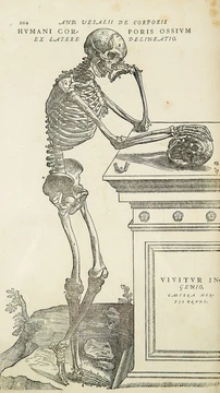
Bladder
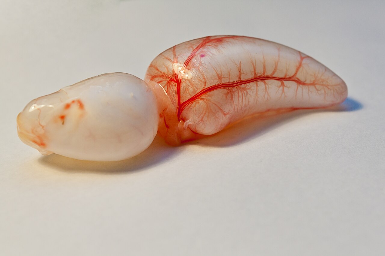
Hollow organ storing urine from the kidneys of vertebrates.

own work by me, Grand-Duc . Die volle Nennung des Autorenpseudonyms und der Zusc · CC BY-SA 3.0 de
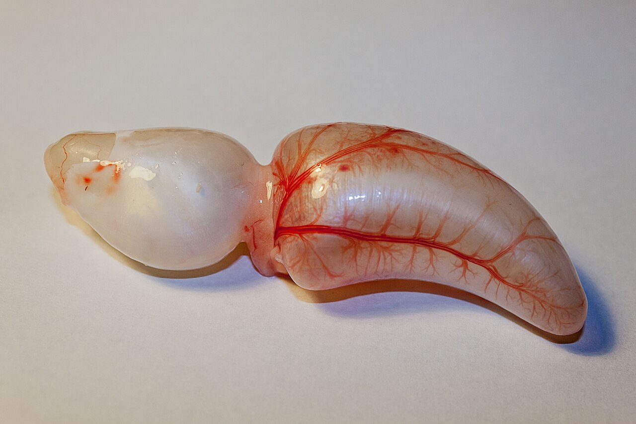
The bladder is a hollow, stretchable organ found in vertebrates that stores urine produced by the kidneys. In humans and other placental mammals, urine flows into the bladder through the ureters and leaves through the urethra. The word "bladder" comes from the Old English *blædre*, meaning 'bladder, blister, or pimple.' Its Latin name is *vesica urinaria*, giving rise to terms like "vesical" and "vesico-" for related structures (e.g., vesical veins). The modern Latin term *cystis* appears in words such as cystitis, which is inflammation of the bladder.
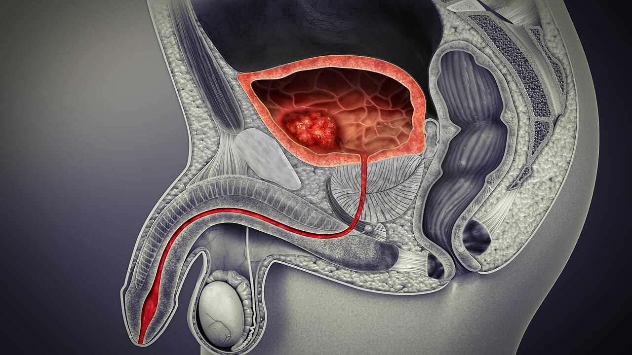
In humans, the bladder is a muscular organ located at the base of the pelvis. Anatomically, it is divided into a broad fundus (base), a body, an apex (or vertex), and a neck. The apex points forward toward the upper part of the pubic symphysis, and from it the median umbilical ligament runs upward along the back of the anterior abdominal wall to the umbilicus. The peritoneum follows this ligament from the apex to the abdominal wall, forming the middle umbilical fold. The neck is the area at the base of the trigone that surrounds the internal urethral orifice, which leads to the urethra. In males, the bladder neck sits next to the prostate gland.
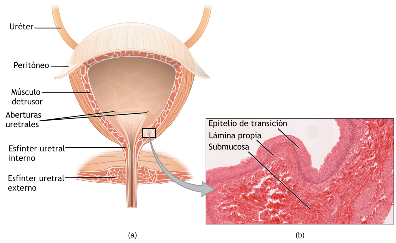
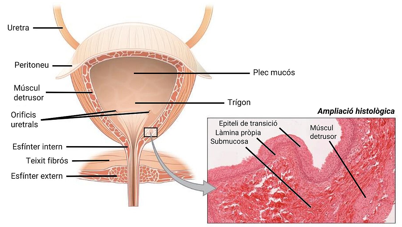
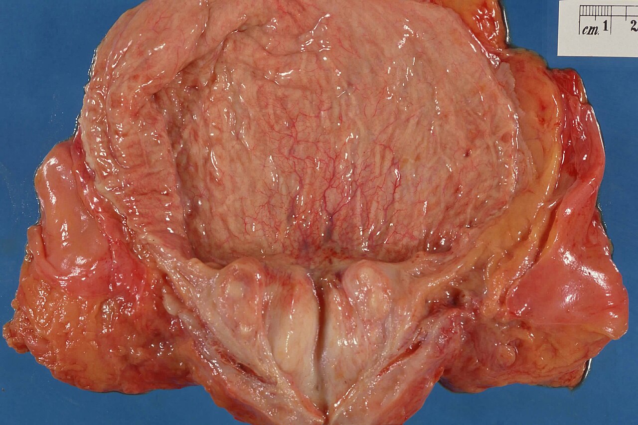
The bladder has three openings: two ureters enter at the ureteric orifices, and the urethra enters at the trigone. Mucosal flaps in front of the ureteric openings act as valves to prevent urine from flowing back into the ureters—a condition called vesicoureteral reflux. Between the two ureteric openings lies a raised area of tissue called the interureteric crest, which forms the upper boundary of the trigone. The trigone is a smooth muscle area that makes up the floor of the bladder above the urethra; its smooth surface allows urine to flow easily into and out of this part, unlike the irregular surface created by the rugae. The bladder wall contains ridges and thick mucosal folds called rugae, which allow the organ to expand. The detrusor muscle, the muscular layer of the wall, consists of smooth muscle fibers arranged in spiral, longitudinal, and circular bundles. It can change length, contract for long periods during voiding, and stay relaxed while the bladder fills. The wall is normally 3–5 mm thick, but when well distended, it is less than 3 mm.
In males, the prostate gland lies outside the urethral opening. The middle lobe of the prostate causes an elevation in the mucous membrane behind the internal urethral orifice, known as the uvula of the urinary bladder. This uvula can enlarge if the prostate becomes enlarged. The bladder sits below the peritoneal cavity near the pelvic floor and behind the pubic symphysis. In males, it lies in front of the rectum, separated by the rectovesical pouch, and is supported by fibers of the levator ani and the prostate gland. In females, it lies in front of the uterus, separated by the vesicouterine pouch, and is supported by the levator ani and the upper part of the vagina.
Blood is supplied to the bladder by the vesical arteries and drained by a network of vesical veins. The superior vesical artery supplies the upper part, and the inferior vesical artery supplies the lower part; both are branches of the internal iliac arteries. In females, the uterine and vaginal arteries provide additional blood supply. Venous drainage starts in a network of small vessels on the lower lateral surfaces of the bladder, which merge and travel with the lateral ligaments of the bladder into the internal iliac veins. Lymph drainage begins in networks throughout the mucosal, muscular, and serosal layers, forming three sets of vessels: one near the trigone draining the bottom, one draining the top, and one draining the outer undersurface. Most of these vessels drain into the external iliac lymph nodes.
The bladder receives both sensory and motor nerve supply from the sympathetic and parasympathetic nervous systems. Motor fibers come from sympathetic fibers (mostly from the superior and inferior hypogastric plexuses and nerves) and from parasympathetic fibers (from the pelvic splanchnic nerves). Sensation related to distension or irritation (e.g., from infection or a stone) is primarily transmitted through the parasympathetic nervous system, traveling via sacral nerves to S2–4, and then to the brain through the dorsal columns of the spinal cord.
Under a microscope, the bladder shows an inner lining (epithelium), three layers of muscle fibers, and an outer adventitia. The inner wall, called the urothelium, is a type of transitional epithelium made of three to six layers of cells; these cells become more cuboidal or flatter depending on whether the bladder is empty or full. The urothelium is lined with a mucous membrane that includes a surface glycocalyx, which protects the underlying cells from urine. The epithelium rests on a thin basement membrane and a lamina propria. This mucosal lining also provides a urothelial barrier against infections. Surrounding these layers are three layers of muscle fibers: an inner layer oriented longitudinally, a middle layer of circular fibers, and an outermost layer of longitudinal fibers—these form the detrusor muscle.
- location
- Base of the pelvis, behind the pubic symphysis
- wall thickness
- 3–5 mm normally; less than 3 mm when well distended
- muscle layer
- Detrusor muscle (smooth muscle fibers in spiral, longitudinal, and circular bundles)
- nerve supply
- Sympathetic and parasympathetic nervous systems
- blood supply
- Superior and inferior vesical arteries (branches of internal iliac arteries)
Lore & Background
The bladder is a hollow muscular organ situated at the base of the pelvis. In gross anatomy, it can be divided into a broad fundus, a body, an apex, and a neck. The apex is directed forward toward the upper part of the pubic symphysis, and from there the median umbilical ligament continues upward on the back of the anterior abdominal wall to the umbilicus. The neck of the bladder surrounds the internal urethral orifice that leads to the urethra. In males, the neck is next to the prostate gland. The bladder has three openings: two ureteric orifices and the urethra at the trigone. The trigone is an area of smooth muscle that forms the floor of the bladder above the urethra. The walls have thick mucosal folds called rugae that allow expansion. The detrusor muscle is the muscular layer of the wall, able to change its length and contract for a long time during voiding while staying relaxed during filling. In the developing embryo, the bladder derives from the urogenital sinus, initially continuous with the allantois. The upper and lower parts develop separately and join around the middle part of development. In infants and young children, the urinary bladder is in the abdomen even when empty.
Reader's Guide
The bladder serves as a critical reservoir for urine, coordinating storage and release through complex neural and muscular mechanisms. Its distensibility, provided by rugae and the detrusor muscle, allows it to hold varying volumes without significant pressure increase. The trigone's smooth tissue facilitates easy urine flow, while mucosal flaps at the ureteric openings prevent backflow. Clinically, the bladder is subject to inflammation (cystitis) and infection, and its proximity to the prostate in males means enlargement of the prostate can affect bladder function. The bladder's development from the urogenital sinus and its anatomical relationships—lying in front of the rectum in males and in front of the uterus in females—underscore its role in pelvic health. Its blood supply via vesical arteries and drainage into internal iliac veins, along with lymphatic drainage to external iliac nodes, support its metabolic needs and immune surveillance.
Did You Know?
- The Latin phrase for 'urinary bladder' is vesica urinaria, and the term vesical or prefix vesico- appear in associated structures such as vesical veins.
- The modern Latin word for 'bladder' – cystis – appears in terms such as cystitis (inflammation of the bladder).
- The detrusor muscle is made of smooth muscle fibers arranged in spiral, longitudinal, and circular bundles.
- In infants and young children, the urinary bladder is in the abdomen even when empty.
Gross Anatomy & Functional Design
The bladder is a hollow muscular organ nestled at the base of the pelvis, resting on the pelvic floor. In gross anatomy it divides into four regions: a broad fundus (base), a body, an apex (or vertex) directed forward toward the upper pubic symphysis, and a neck that surrounds the internal urethral orifice. Three openings punctuate the organ: two ureteric orifices through which urine enters from the kidneys, and the urethral opening at the trigone through which urine exits. The trigone is a smooth-muscle floor above the urethra, bounded superiorly by the interureteric crest. Mucosal flaps positioned before each ureteric orifice act as one-way valves, preventing vesicoureteral reflux. The inner walls bear thick mucosal folds called rugae that permit expansion. The detrusor muscle—composed of smooth muscle arranged in spiral, longitudinal, and circular bundles—remains relaxed during filling and contracts over extended periods during voiding. The wall measures 3–5 mm thick at rest and thins below 3 mm when well distended.
Microscopic Architecture & Protective Layers
Under the microscope, the bladder reveals a layered construction designed for both protection and flexibility. The innermost lining is the urothelium, a transitional epithelium of three to six cell layers that shift shape—becoming more cuboidal or flatter—depending on whether the organ is empty or full. A surface glycocalyx coats these cells, shielding them from the chemical assault of urine, while a thin basement membrane and lamina propria anchor the epithelium to the tissue beneath. This mucosal lining also functions as a urothelial barrier against the passage of infections. Beneath the epithelium lie three distinct muscle-fibre layers: an inner longitudinal layer, a middle circular layer, and an outer longitudinal layer, together forming the detrusor muscle visible to the naked eye. The entire structure is wrapped externally by a serous membrane called the adventitia, which provides a protective outer covering for the organ.
Vascular, Lymphatic & Neural Wiring
The bladder's blood supply arrives via the vesical arteries, both of which are branches of the internal iliac arteries. The superior vesical artery feeds the upper portion, while the inferior vesical artery serves the lower portion; in females, the uterine and vaginal arteries contribute additional flow. Venous drainage begins in small vessel networks on the lower lateral surfaces, coalescing and traveling with the lateral ligaments into the internal iliac veins. Lymphatic drainage is organized into three vessel sets—one near the trigone, one draining the top, and one from the outer undersurface—most of which empty into the external iliac lymph nodes. Neurologically, the organ receives both sympathetic and parasympathetic input. Motor signals arrive via the superior and inferior hypogastric plexuses (sympathetic) and the pelvic splanchnic nerves (parasympathetic). Sensory information about distension or irritation, such as from infection or a stone, travels primarily through parasympathetic pathways via sacral nerves S2–S4, then ascends through the dorsal columns of the spinal cord to the brain.
Embryonic Origins & Sex-Specific Relationships
In the developing embryo, the bladder's lineage traces back to the cloaca at the hind end. Between the fourth and seventh weeks, a wall called the urorectal septum divides the cloaca into a urogenital sinus and the beginnings of the anal canal. The urogenital sinus then subdivides into three parts, with the upper portion giving rise to the bladder. In the adult, the organ's neighbors differ by sex. In males, the prostate gland sits outside the urethral opening, and its middle lobe creates a mucosal elevation called the uvula of the urinary bladder, which can enlarge alongside prostatic growth. The bladder lies anterior to the rectum, separated by the rectovesical pouch, and is supported by levator ani fibres and the prostate. In females, the bladder sits in front of the uterus, separated by the vesicouterine pouch, with support from the levator ani and the upper vagina. The apex connects via the median umbilical ligament to the umbilicus, and the peritoneum drapes from the apex to form the middle umbilical fold on the anterior abdominal wall.
Gallery
Frequently Asked Questions
What is the Bladder in animal anatomy?
The Bladder is a flexible, hollow sac found in all vertebrates whose job is to hold urine produced by the kidneys. It can expand and contract as needed to accommodate varying volumes of fluid.
Where exactly is the Bladder situated in the body?
It sits at the very bottom of the pelvic cavity, tucked behind the pubic bone (pubic symphysis). This low position keeps it protected by the bony pelvis.
What muscle is responsible for emptying the Bladder?
A smooth muscle called the detrusor forms the Bladder's muscular wall, arranged in spiral, longitudinal, and circular fiber bundles. When these fibers contract together, they squeeze urine out through the urethra.
How does urine get into and out of the Bladder?
Two ureter tubes carry urine down from the kidneys into the Bladder, and a single urethral channel serves as the exit route. In placental mammals like humans, this in-and-out pathway is the standard arrangement.
What keeps the Bladder alive—blood and nerve supply?
It receives its blood from the superior and inferior vesical arteries, which branch off the internal iliac arteries. Both sympathetic and parasympathetic nerves innervate the organ, coordinating storage and release.
More in Animal Anatomy 1-17
Spotted an error? Know more?
This is a living reference — every entry is fact-audited, and reader corrections feed straight into our audit queue. Suggest an edit · See this site's audit record